 Movie
Movie Controller

[English] 日本語
 Yorodumi
- PDB-1qk1: CRYSTAL STRUCTURE OF HUMAN UBIQUITOUS MITOCHONDRIAL CREATINE KINASE -
+ Open data

- Basic information
| Entry | Database: PDB / ID: 1qk1 | ||||||
|---|---|---|---|---|---|---|---|
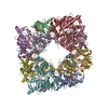
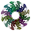
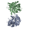
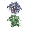
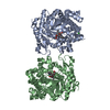
| Title | CRYSTAL STRUCTURE OF HUMAN UBIQUITOUS MITOCHONDRIAL CREATINE KINASE | ||||||
 Components Components | CREATINE KINASE, UBIQUITOUS MITOCHONDRIAL | ||||||
 Keywords Keywords | TRANSFERASE (CREATINE KINASE) / MITOCHONDRIAL CREATINE KINASE / CANCER / CELLULAR ENERGY METABOLISM / GUANIDINO KINASE / MITOCHONDRIAL PERMEABILITY TRANSITION / OCTAMER STABILITY | ||||||
| Function / homology |  Function and homology information Function and homology informationcreatine kinase / phosphocreatine biosynthetic process / Creatine metabolism / creatine kinase activity / mitochondrial inner membrane / mitochondrion / ATP binding Similarity search - Function | ||||||
| Biological species |  HOMO SAPIENS (human) HOMO SAPIENS (human) | ||||||
| Method |  X-RAY DIFFRACTION / MOLECULAR REPLACEMENT / Resolution: 2.7 Å X-RAY DIFFRACTION / MOLECULAR REPLACEMENT / Resolution: 2.7 Å | ||||||
 Authors Authors | Eder, M. / Schlattner, U. / Fritz-Wolf, K. / Wallimann, T. / Kabsch, W. | ||||||
 Citation Citation | Journal: Proteins: Struct.,Funct., Genet. / Year: 2000 Title: Crystal Structure of Human Ubiquitous Mitochondrial Creatine Kinase Authors: Eder, M. / Fritz-Wolf, K. / Kabsch, W. / Wallimann, T. / Schlattner, U. #1: Journal: Nature / Year: 1996Title: Structure of Mitochondrial Creatine Kinase: Authors: Fritz-Wolf, K. / Schnyder, T. / Wallimann, T. / Kabsch, W. | ||||||
| History |
|
- Structure visualization
| Structure viewer | Molecule: MolmilJmol/JSmol |
|---|
- Downloads & links
-Download
| PDBx/mmCIF format | 1qk1.cif.gz | 583.2 KB | Display | PDBx/mmCIF format |
|---|---|---|---|---|
| PDB format | pdb1qk1.ent.gz | 489 KB | Display | PDB format |
| PDBx/mmJSON format | 1qk1.json.gz | Tree view | PDBx/mmJSON format | |
| Others |  Other downloads Other downloads |
-Validation report
| Arichive directory | https://data.pdbj.org/pub/pdb/validation_reports/qk/1qk1ftp://data.pdbj.org/pub/pdb/validation_reports/qk/1qk1 | HTTPS FTP |
|---|
-Related structure data
| Related structure data | 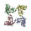 1crkS S: Starting model for refinement |
|---|---|
| Similar structure data |

-Links
 PDBj
PDBj

- Assembly
| Deposited unit | 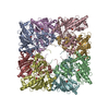
| ||||||||
|---|---|---|---|---|---|---|---|---|---|
| 1 |
| ||||||||
| Unit cell |
| ||||||||
| Details | THE MOLECULE EXISTS AS AN OCTAMER COMPOSED OFFOUR UMTCK HOMODIMERS. |
-Components
| #1: Protein | Mass: 43216.086 Da / Num. of mol.: 8 Source method: isolated from a genetically manipulated source Details: RECOMBINANT HUMAN UMTCK HAS AN ADDITIONAL ALA AT THE N-TERMINUS Source: (gene. exp.) HOMO SAPIENS (human) / Tissue: PLACENTA / Cellular location: MITOCHONDRIAL INNER MEMBRANEGene: GENBANK ACCESSION J04469 GENE: GENBANK ACCESSION J04469 Plasmid: PUS04 / Production host:  #2: Chemical | ChemComp-PO4 /   Mass: 94.971 Da / Num. of mol.: 8 / Source method: obtained synthetically / Formula: PO4 Mass: 94.971 Da / Num. of mol.: 8 / Source method: obtained synthetically / Formula: PO4#3: Water | ChemComp-HOH / |  Mass: 18.015 Da / Num. of mol.: 293 / Source method: isolated from a natural source / Formula: H2O Mass: 18.015 Da / Num. of mol.: 293 / Source method: isolated from a natural source / Formula: H2OSource details | FOR THE MIA-CK STUDIED HERE THE MATURE PROTEIN SEQUENCE STARTS AT POSITION 39 OF THE PRECURSOR ...FOR THE MIA-CK STUDIED HERE THE MATURE PROTEIN SEQUENCE STARTS AT POSITION 39 OF THE PRECURSOR PROTEIN. THE SWS P12532 STARTS AT 40, WITH RESIDUES 1 39 MITOCHONDR | |
|---|
-Experimental details
-Experiment
| Experiment | Method: X-RAY DIFFRACTION / Number of used crystals: 5 |
|---|
- Sample preparation
| Crystal | Density Matthews: 3.5 Å3/Da / Density % sol: 65 % | |||||||||||||||||||||||||
|---|---|---|---|---|---|---|---|---|---|---|---|---|---|---|---|---|---|---|---|---|---|---|---|---|---|---|
| Crystal grow | pH: 6.75 Details: 0.025M NA-PHOSPHATE, 3 MM DTT, PH 6.75, 5 MG/ML PROTEIN | |||||||||||||||||||||||||
| Crystal | *PLUS Density % sol: 65 % | |||||||||||||||||||||||||
| Crystal grow | *PLUS Temperature: 4 ℃ / Method: batch method | |||||||||||||||||||||||||
| Components of the solutions | *PLUS
|
-Data collection
| Diffraction | Mean temperature: 277 K |
|---|---|
| Diffraction source | Source: ROTATING ANODE / Type: ELLIOTT GX-18 / Wavelength: 1.5418 |
| Detector | Type: MULTIWIRE SIEMENS / Detector: AREA DETECTOR / Date: Dec 15, 1998 / Details: FRANKS DOUBLE MIRROR |
| Radiation | Protocol: SINGLE WAVELENGTH / Monochromatic (M) / Laue (L): M / Scattering type: x-ray |
| Radiation wavelength | Wavelength: 1.5418 Å / Relative weight: 1 |
| Reflection | Resolution: 2.7→50 Å / Num. obs: 130945 / % possible obs: 99.4 % / Redundancy: 4.7 % / Biso Wilson estimate: 49.3 Å2 / Rmerge(I) obs: 0.092 / Net I/σ(I): 11.1 |
| Reflection shell | Resolution: 2.7→2.8 Å / Redundancy: 3.2 % / Rmerge(I) obs: 0.313 / Mean I/σ(I) obs: 2.7 / % possible all: 99.1 |
| Reflection | *PLUS Num. measured all: 616668 |
| Reflection shell | *PLUS % possible obs: 99.1 % / Num. unique obs: 13429 / Num. measured obs: 43256 |
- Processing
| Software |
| ||||||||||||||||||||||||||||||||||||||||||||||||||||||||||||||||||||||||||||||||
|---|---|---|---|---|---|---|---|---|---|---|---|---|---|---|---|---|---|---|---|---|---|---|---|---|---|---|---|---|---|---|---|---|---|---|---|---|---|---|---|---|---|---|---|---|---|---|---|---|---|---|---|---|---|---|---|---|---|---|---|---|---|---|---|---|---|---|---|---|---|---|---|---|---|---|---|---|---|---|---|---|---|
| Refinement | Method to determine structure: MOLECULAR REPLACEMENT Starting model: 1CRK, SARCOMERIC MITOCHONDRIAL CREATINE KINASE Resolution: 2.7→50 Å / Rfactor Rfree error: 0.003 / Data cutoff high absF: 10000 / Isotropic thermal model: RESTRAINED / Cross valid method: FREE R-VALUE / σ(F): 0 Details: DISORDERED RESIDUES 316-325 AND 375- 379 (CHAIN A-H) WERE MODELED
| ||||||||||||||||||||||||||||||||||||||||||||||||||||||||||||||||||||||||||||||||
| Solvent computation | Solvent model: FLAT MODEL / Bsol: 23.97 Å2 / ksol: 0.2798 e/Å3 | ||||||||||||||||||||||||||||||||||||||||||||||||||||||||||||||||||||||||||||||||
| Displacement parameters | Biso mean: 45.3 Å2
| ||||||||||||||||||||||||||||||||||||||||||||||||||||||||||||||||||||||||||||||||
| Refine analyze |
| ||||||||||||||||||||||||||||||||||||||||||||||||||||||||||||||||||||||||||||||||
| Refinement step | Cycle: LAST / Resolution: 2.7→50 Å
| ||||||||||||||||||||||||||||||||||||||||||||||||||||||||||||||||||||||||||||||||
| Refine LS restraints |
| ||||||||||||||||||||||||||||||||||||||||||||||||||||||||||||||||||||||||||||||||
| LS refinement shell | Resolution: 2.7→2.8 Å / Rfactor Rfree error: 0.012 / Total num. of bins used: 10
| ||||||||||||||||||||||||||||||||||||||||||||||||||||||||||||||||||||||||||||||||
| Xplor file |
| ||||||||||||||||||||||||||||||||||||||||||||||||||||||||||||||||||||||||||||||||
| Software | *PLUS Name: CNS / Version: 0.5 / Classification: refinement | ||||||||||||||||||||||||||||||||||||||||||||||||||||||||||||||||||||||||||||||||
| Refinement | *PLUS Lowest resolution: 50 Å | ||||||||||||||||||||||||||||||||||||||||||||||||||||||||||||||||||||||||||||||||
| Solvent computation | *PLUS | ||||||||||||||||||||||||||||||||||||||||||||||||||||||||||||||||||||||||||||||||
| Displacement parameters | *PLUS | ||||||||||||||||||||||||||||||||||||||||||||||||||||||||||||||||||||||||||||||||
| Refine LS restraints | *PLUS
|
