 Movie
Movie Controller

[English] 日本語
 Yorodumi
- PDB-1qi6: SECOND APO FORM OF AN NADP DEPENDENT ALDEHYDE DEHYDROGENASE WITH ... -
+ Open data

- Basic information
| Entry | Database: PDB / ID: 1qi6 | ||||||
|---|---|---|---|---|---|---|---|
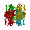
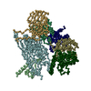
| Title | SECOND APO FORM OF AN NADP DEPENDENT ALDEHYDE DEHYDROGENASE WITH GLU250 SITUATED 3.7 A FROM CYS284 | ||||||
 Components Components | PROTEIN (NADP DEPENDENT NONPHOSPHORYLATING GLYCERALDEHYDE-3-PHOSPHATE DEHYDROGENASE) | ||||||
 Keywords Keywords | OXIDOREDUCTASE | ||||||
| Function / homology |  Function and homology information Function and homology informationglyceraldehyde-3-phosphate dehydrogenase (NADP+) / glyceraldehyde-3-phosphate dehydrogenase (NADP+) (non-phosphorylating) activity / lactaldehyde dehydrogenase (NAD+) activity / glyceraldehyde-3-phosphate dehydrogenase (NADP+) (phosphorylating) activity Similarity search - Function | ||||||
| Biological species |  Streptococcus mutans (bacteria) Streptococcus mutans (bacteria) | ||||||
| Method |  X-RAY DIFFRACTION / MOLECULAR REPLACEMENT / Resolution: 2.5 Å X-RAY DIFFRACTION / MOLECULAR REPLACEMENT / Resolution: 2.5 Å | ||||||
 Authors Authors | Cobessi, D. / Tete-Favier, F. / Marchal, S. / Branlant, G. / Aubry, A. | ||||||
 Citation Citation | Journal: J.Mol.Biol. / Year: 2000 Title: Structural and biochemical investigations of the catalytic mechanism of an NADP-dependent aldehyde dehydrogenase from Streptococcus mutans. Authors: Cobessi, D. / Tete-Favier, F. / Marchal, S. / Branlant, G. / Aubry, A. | ||||||
| History |
|
- Structure visualization
| Structure viewer | Molecule: MolmilJmol/JSmol |
|---|
- Downloads & links
-Download
| PDBx/mmCIF format | 1qi6.cif.gz | 362 KB | Display | PDBx/mmCIF format |
|---|---|---|---|---|
| PDB format | pdb1qi6.ent.gz | 295.4 KB | Display | PDB format |
| PDBx/mmJSON format | 1qi6.json.gz | Tree view | PDBx/mmJSON format | |
| Others |  Other downloads Other downloads |
-Validation report
| Arichive directory | https://data.pdbj.org/pub/pdb/validation_reports/qi/1qi6ftp://data.pdbj.org/pub/pdb/validation_reports/qi/1qi6 | HTTPS FTP |
|---|
-Related structure data
| Related structure data | 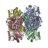 1qi1C  1euhS S: Starting model for refinement C: citing same article ( |
|---|---|
| Similar structure data |


-Links
 PDBj
PDBj

- Assembly
| Deposited unit | 
| ||||||||||||||||
|---|---|---|---|---|---|---|---|---|---|---|---|---|---|---|---|---|---|
| 1 |
| ||||||||||||||||
| Unit cell |
| ||||||||||||||||
| Noncrystallographic symmetry (NCS) | NCS oper:
|
-Components
| #1: Protein | Mass: 51276.438 Da / Num. of mol.: 4 Source method: isolated from a genetically manipulated source Details: SO4 / Source: (gene. exp.) Streptococcus mutans (bacteria) / Production host: References: UniProt: Q59931, glyceraldehyde-3-phosphate dehydrogenase (NADP+) #2: Chemical | ChemComp-SO4 /   Mass: 96.063 Da / Num. of mol.: 12 / Source method: obtained synthetically / Formula: SO4 Mass: 96.063 Da / Num. of mol.: 12 / Source method: obtained synthetically / Formula: SO4#3: Water | ChemComp-HOH / |  Mass: 18.015 Da / Num. of mol.: 565 / Source method: isolated from a natural source / Formula: H2O Mass: 18.015 Da / Num. of mol.: 565 / Source method: isolated from a natural source / Formula: H2O |
|---|
-Experimental details
-Experiment
| Experiment | Method: X-RAY DIFFRACTION / Number of used crystals: 1 |
|---|
- Sample preparation
| Crystal | Density Matthews: 2.98 Å3/Da / Density % sol: 60 % | |||||||||||||||||||||||||
|---|---|---|---|---|---|---|---|---|---|---|---|---|---|---|---|---|---|---|---|---|---|---|---|---|---|---|
| Crystal grow | pH: 8.5 Details: 2.0 M AMMONIUM SULPHATE, 2 % PEG 400, HEPES O.1 M PH 7.5, PROTEIN 5MG/ML, pH 8.5 | |||||||||||||||||||||||||
| Crystal grow | *PLUS Temperature: 20 ℃ / pH: 7.5 / Method: vapor diffusion, hanging drop | |||||||||||||||||||||||||
| Components of the solutions | *PLUS
|
-Data collection
| Diffraction | Mean temperature: 100 K |
|---|---|
| Diffraction source | Source: ROTATING ANODE / Type: ENRAF-NONIUS / Wavelength: 1.5418 |
| Detector | Type: ENRAF-NONIUS / Detector: IMAGE PLATE / Date: Jun 15, 1997 / Details: MIRRORS |
| Radiation | Monochromator: NI FILTER / Protocol: SINGLE WAVELENGTH / Monochromatic (M) / Laue (L): M / Scattering type: x-ray |
| Radiation wavelength | Wavelength: 1.5418 Å / Relative weight: 1 |
| Reflection | Resolution: 2.5→20 Å / Num. obs: 81791 / % possible obs: 96 % / Redundancy: 4.5 % / Rmerge(I) obs: 0.059 / Rsym value: 0.059 |
| Reflection shell | Resolution: 2.5→2.59 Å / Rmerge(I) obs: 0.188 / Rsym value: 0.188 / % possible all: 68.4 |
- Processing
| Software |
| ||||||||||||||||||||||||||||||||||||||||||||||||||||||||||||
|---|---|---|---|---|---|---|---|---|---|---|---|---|---|---|---|---|---|---|---|---|---|---|---|---|---|---|---|---|---|---|---|---|---|---|---|---|---|---|---|---|---|---|---|---|---|---|---|---|---|---|---|---|---|---|---|---|---|---|---|---|---|
| Refinement | Method to determine structure: MOLECULAR REPLACEMENT Starting model: 1EUH Resolution: 2.5→8 Å / Data cutoff high absF: 3500 / Data cutoff low absF: 0.1 / Cross valid method: THROUGHOUT / σ(F): 0
| ||||||||||||||||||||||||||||||||||||||||||||||||||||||||||||
| Refinement step | Cycle: LAST / Resolution: 2.5→8 Å
| ||||||||||||||||||||||||||||||||||||||||||||||||||||||||||||
| Refine LS restraints |
| ||||||||||||||||||||||||||||||||||||||||||||||||||||||||||||
| Software | *PLUS Name: X-PLOR / Version: 3.8 / Classification: refinement | ||||||||||||||||||||||||||||||||||||||||||||||||||||||||||||
| Refinement | *PLUS Highest resolution: 2.5 Å / σ(F): 0 / Rfactor Rfree: 0.28 | ||||||||||||||||||||||||||||||||||||||||||||||||||||||||||||
| Solvent computation | *PLUS | ||||||||||||||||||||||||||||||||||||||||||||||||||||||||||||
| Displacement parameters | *PLUS | ||||||||||||||||||||||||||||||||||||||||||||||||||||||||||||
| Refine LS restraints | *PLUS
|
