 Movie
Movie Controller


+ Open data

- Basic information
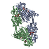
| Entry | Database: PDB / ID: 1qha | ||||||
|---|---|---|---|---|---|---|---|
| Title | HUMAN HEXOKINASE TYPE I COMPLEXED WITH ATP ANALOGUE AMP-PNP | ||||||
 Components Components | PROTEIN (HEXOKINASE) | ||||||
 Keywords Keywords | TRANSFERASE / KINASE / GLYCOLYSIS / PHOSPHOTRANSFERASE | ||||||
| Function / homology |  Function and homology information Function and homology informationDefective HK1 causes hexokinase deficiency (HK deficiency) / glucosamine kinase activity / : / hexokinase activity / Synthesis of GDP-mannose / carbohydrate phosphorylation / mannokinase activity / maintenance of protein location in mitochondrion / hexokinase / : ...Defective HK1 causes hexokinase deficiency (HK deficiency) / glucosamine kinase activity / : / hexokinase activity / Synthesis of GDP-mannose / carbohydrate phosphorylation / mannokinase activity / maintenance of protein location in mitochondrion / hexokinase / : / GDP-mannose biosynthetic process / fructokinase activity / glucokinase activity / mannose metabolic process / positive regulation of cytokine production involved in immune response / glucose 6-phosphate metabolic process / peptidoglycan binding / D-glucose binding / fructose 6-phosphate metabolic process / Glycolysis / canonical glycolysis / intracellular glucose homeostasis / glycolytic process / positive regulation of interleukin-1 beta production / glucose metabolic process / mitochondrial outer membrane / membrane raft / inflammatory response / innate immune response / mitochondrion / ATP binding / cytosol Similarity search - Function | ||||||
| Biological species |  Homo sapiens (human) Homo sapiens (human) | ||||||
| Method |  X-RAY DIFFRACTION / SYNCHROTRON / MOLECULAR REPLACEMENT / Resolution: 2.25 Å X-RAY DIFFRACTION / SYNCHROTRON / MOLECULAR REPLACEMENT / Resolution: 2.25 Å | ||||||
 Authors Authors | Rosano, C. / Sabini, E. / Deriu, D. / Magnani, M. / Bolognesi, M. | ||||||
 Citation Citation | Journal: Structure Fold.Des. / Year: 1999 Title: Binding of non-catalytic ATP to human hexokinase I highlights the structural components for enzyme-membrane association control. Authors: Rosano, C. / Sabini, E. / Rizzi, M. / Deriu, D. / Murshudov, G. / Bianchi, M. / Serafini, G. / Magnani, M. / Bolognesi, M. #1: Journal: Protein Pept.Lett. / Year: 1998Title: Expression, Purification and Preliminary Crystallographic Studies of Human Hexokinase I Authors: Sabini, E. / Rosano, C. / Capasso, C. / Rizzi, M. / Bolognesi, M. / Bianchi, M. / Serafini, G. / Bartolucci, E. / Magnani, M. | ||||||
| History |
|
- Structure visualization
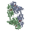
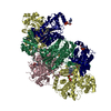
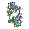
| Structure viewer | Molecule: MolmilJmol/JSmol |
|---|
- Downloads & links
-Download
| PDBx/mmCIF format | 1qha.cif.gz | 375.8 KB | Display | PDBx/mmCIF format |
|---|---|---|---|---|
| PDB format | pdb1qha.ent.gz | 298.4 KB | Display | PDB format |
| PDBx/mmJSON format | 1qha.json.gz | Tree view | PDBx/mmJSON format | |
| Others |  Other downloads Other downloads |
-Validation report
| Arichive directory | https://data.pdbj.org/pub/pdb/validation_reports/qh/1qhaftp://data.pdbj.org/pub/pdb/validation_reports/qh/1qha | HTTPS FTP |
|---|
-Related structure data
| Related structure data | 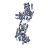 1hkbS S: Starting model for refinement |
|---|---|
| Similar structure data |


-Links
 PDBj
PDBj


- Assembly
| Deposited unit | 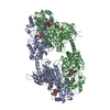
| ||||||||
|---|---|---|---|---|---|---|---|---|---|
| 1 |
| ||||||||
| Unit cell |
|
-Components
-Protein , 1 types, 2 molecules AB
| #1: Protein | Mass: 102624.055 Da / Num. of mol.: 2 Source method: isolated from a genetically manipulated source Details: COMPLEXED WITH ATP ANALOGUE AMP-PNP / Source: (gene. exp.) Homo sapiens (human) / Production host:  |
|---|
-Sugars , 2 types, 8 molecules 
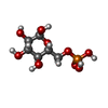
| #2: Sugar | ChemComp-GLC /  Type: D-saccharide, alpha linking / Mass: 180.156 Da / Num. of mol.: 4 / Source method: isolated from a natural source / Formula: C6H12O6 Type: D-saccharide, alpha linking / Mass: 180.156 Da / Num. of mol.: 4 / Source method: isolated from a natural source / Formula: C6H12O6#3: Sugar | ChemComp-G6P /  Type: D-saccharide, alpha linking / Mass: 260.136 Da / Num. of mol.: 4 / Source method: isolated from a natural source / Formula: C6H13O9P Type: D-saccharide, alpha linking / Mass: 260.136 Da / Num. of mol.: 4 / Source method: isolated from a natural source / Formula: C6H13O9P |
|---|
-Non-polymers , 3 types, 468 molecules 


| #4: Chemical | ChemComp-MG /  Mass: 24.305 Da / Num. of mol.: 6 / Source method: obtained synthetically / Formula: Mg Mass: 24.305 Da / Num. of mol.: 6 / Source method: obtained synthetically / Formula: Mg#5: Chemical |  Mass: 506.196 Da / Num. of mol.: 2 / Source method: obtained synthetically / Formula: C10H17N6O12P3 / Comment: AMP-PNP, energy-carrying molecule analogue*YM Mass: 506.196 Da / Num. of mol.: 2 / Source method: obtained synthetically / Formula: C10H17N6O12P3 / Comment: AMP-PNP, energy-carrying molecule analogue*YM#6: Water | ChemComp-HOH / | Mass: 18.015 Da / Num. of mol.: 460 / Source method: isolated from a natural source / Formula: H2O |
|---|
-Details
| Nonpolymer details | 4 MOLECULES IN ALPHA-ANOMER CONFORMATI |
|---|
-Experimental details
-Experiment
| Experiment | Method: X-RAY DIFFRACTION / Number of used crystals: 1 |
|---|
- Sample preparation
| Crystal | Density Matthews: 3.11 Å3/Da / Density % sol: 60.47 % | |||||||||||||||||||||||||||||||||||||||||||||||||||||||||||||||||||||||||||||||||||||||||||
|---|---|---|---|---|---|---|---|---|---|---|---|---|---|---|---|---|---|---|---|---|---|---|---|---|---|---|---|---|---|---|---|---|---|---|---|---|---|---|---|---|---|---|---|---|---|---|---|---|---|---|---|---|---|---|---|---|---|---|---|---|---|---|---|---|---|---|---|---|---|---|---|---|---|---|---|---|---|---|---|---|---|---|---|---|---|---|---|---|---|---|---|---|
| Crystal grow | pH: 6.5 / Details: pH 6.5 | |||||||||||||||||||||||||||||||||||||||||||||||||||||||||||||||||||||||||||||||||||||||||||
| Crystal grow | *PLUS Temperature: 4 ℃ / pH: 5.5 / Method: vapor diffusion, hanging dropDetails: drop contained 0.004ml solutionA, 0.001ml reservoir solution and 0.001ml solutionB. | |||||||||||||||||||||||||||||||||||||||||||||||||||||||||||||||||||||||||||||||||||||||||||
| Components of the solutions | *PLUS
|
-Data collection
| Diffraction | Mean temperature: 100 K |
|---|---|
| Diffraction source | Source: SYNCHROTRON / Site: ESRF  / Type: ESRF / Wavelength: 0.936 / Type: ESRF / Wavelength: 0.936 |
| Detector | Type: MARRESEARCH / Detector: CCD / Date: Feb 1, 1999 |
| Radiation | Protocol: SINGLE WAVELENGTH / Monochromatic (M) / Laue (L): M / Scattering type: x-ray |
| Radiation wavelength | Wavelength: 0.936 Å / Relative weight: 1 |
| Reflection | Resolution: 2.25→38 Å / Num. obs: 102729 / % possible obs: 86.6 % / Redundancy: 4 % / Rmerge(I) obs: 0.05 / Rsym value: 0.097 / Net I/σ(I): 8 |
| Reflection shell | Resolution: 2.25→2.3 Å / Mean I/σ(I) obs: 6.8 |
| Reflection | *PLUS Num. measured all: 405442 / Rmerge(I) obs: 0.05 |
- Processing
| Software |
| |||||||||||||||||||||||||||||||||||||||||||||||||||||||||||||||
|---|---|---|---|---|---|---|---|---|---|---|---|---|---|---|---|---|---|---|---|---|---|---|---|---|---|---|---|---|---|---|---|---|---|---|---|---|---|---|---|---|---|---|---|---|---|---|---|---|---|---|---|---|---|---|---|---|---|---|---|---|---|---|---|---|
| Refinement | Method to determine structure: MOLECULAR REPLACEMENT Starting model: 1HKB Resolution: 2.25→12 Å / σ(F): 0
| |||||||||||||||||||||||||||||||||||||||||||||||||||||||||||||||
| Refinement step | Cycle: LAST / Resolution: 2.25→12 Å
| |||||||||||||||||||||||||||||||||||||||||||||||||||||||||||||||
| Refine LS restraints |
| |||||||||||||||||||||||||||||||||||||||||||||||||||||||||||||||
| Software | *PLUS Name: REFMAC / Classification: refinement | |||||||||||||||||||||||||||||||||||||||||||||||||||||||||||||||
| Refinement | *PLUS Lowest resolution: 12 Å / σ(F): 0 / % reflection Rfree: 5 % / Rfactor obs: 0.208 | |||||||||||||||||||||||||||||||||||||||||||||||||||||||||||||||
| Solvent computation | *PLUS | |||||||||||||||||||||||||||||||||||||||||||||||||||||||||||||||
| Displacement parameters | *PLUS |
