 Movie
Movie Controller

[English] 日本語
 Yorodumi
- PDB-1qcr: CRYSTAL STRUCTURE OF BOVINE MITOCHONDRIAL CYTOCHROME BC1 COMPLEX,... -
+ Open data

- Basic information
| Entry | Database: PDB / ID: 1qcr | ||||||
|---|---|---|---|---|---|---|---|
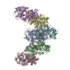
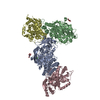
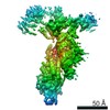
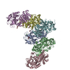
| Title | CRYSTAL STRUCTURE OF BOVINE MITOCHONDRIAL CYTOCHROME BC1 COMPLEX, ALPHA CARBON ATOMS ONLY | ||||||
 Components Components | (UBIQUINOL CYTOCHROME C ...) x 11 | ||||||
 Keywords Keywords | OXIDOREDUCTASE / BC1 / QCR / MEMBRANE PROTEIN / PROTON TRANSLOCATION / ELECTRON TRANSFER / PROTEASE / MPP / MITOCHONDRIAL PROCESSING PEPTIDASE / CYTOCHROME C1 / CYTOCHROME B / RIESKE / IRON SULFER PROTEIN | ||||||
| Function / homology |  Function and homology information Function and homology informationComplex III assembly / subthalamus development / pons development / cerebellar Purkinje cell layer development / Respiratory electron transport / pyramidal neuron development / thalamus development / Mitochondrial translation termination / respiratory chain complex III / quinol-cytochrome-c reductase ...Complex III assembly / subthalamus development / pons development / cerebellar Purkinje cell layer development / Respiratory electron transport / pyramidal neuron development / thalamus development / Mitochondrial translation termination / respiratory chain complex III / quinol-cytochrome-c reductase / quinol-cytochrome-c reductase activity / mitochondrial electron transport, ubiquinol to cytochrome c / Mitochondrial protein degradation / hypothalamus development / midbrain development / ubiquinone binding / respiratory electron transport chain / hippocampus development / metalloendopeptidase activity / 2 iron, 2 sulfur cluster binding / mitochondrial membrane / oxidoreductase activity / mitochondrial inner membrane / heme binding / protein-containing complex / mitochondrion / proteolysis / membrane / metal ion binding Similarity search - Function | ||||||
| Biological species |  | ||||||
| Method |  X-RAY DIFFRACTION / SYNCHROTRON / MIR / Resolution: 2.7 Å X-RAY DIFFRACTION / SYNCHROTRON / MIR / Resolution: 2.7 Å | ||||||
 Authors Authors | Xia, D. / Yu, C.A. / Kim, H. / Xia, J.Z. / Kachurin, A. / Zhang, L. / Yu, L. / Deisenhofer, J. | ||||||
 Citation Citation | Journal: Science / Year: 1997 Title: Crystal structure of the cytochrome bc1 complex from bovine heart mitochondria. Authors: Xia, D. / Yu, C.A. / Kim, H. / Xia, J.Z. / Kachurin, A.M. / Zhang, L. / Yu, L. / Deisenhofer, J. #1: Journal: Science / Year: 1997Title: Erratum. Crystal Structure of the Cytochrome Bc1 Complex from Bovine Heart Mitochondria Authors: Xia, D. / Yu, C.A. / Kim, H. / Xia, J.Z. / Kachurin, A.M. / Zhang, L. / Yu, L. / Deisenhofer, J. | ||||||
| History |
|
- Structure visualization
| Structure viewer | Molecule: MolmilJmol/JSmol |
|---|
- Downloads & links
-Download
| PDBx/mmCIF format | 1qcr.cif.gz | 84.9 KB | Display | PDBx/mmCIF format |
|---|---|---|---|---|
| PDB format | pdb1qcr.ent.gz | 47.5 KB | Display | PDB format |
| PDBx/mmJSON format | 1qcr.json.gz | Tree view | PDBx/mmJSON format | |
| Others |  Other downloads Other downloads |
-Validation report
| Arichive directory | https://data.pdbj.org/pub/pdb/validation_reports/qc/1qcrftp://data.pdbj.org/pub/pdb/validation_reports/qc/1qcr | HTTPS FTP |
|---|
-Related structure data
| Similar structure data |
|---|


-Links
 PDBj
PDBj


- Assembly
| Deposited unit | 
| ||||||||
|---|---|---|---|---|---|---|---|---|---|
| 1 |
| ||||||||
| Unit cell |
|
-Components
-UBIQUINOL CYTOCHROME C ... , 11 types, 11 molecules ABCDEFGHIJK
| #1: Protein | Mass: 49252.227 Da / Num. of mol.: 1 / Source method: isolated from a natural source / Source: (natural) |
|---|---|
| #2: Protein | Mass: 45023.621 Da / Num. of mol.: 1 / Source method: isolated from a natural source / Source: (natural) |
| #3: Protein | Mass: 42489.145 Da / Num. of mol.: 1 / Source method: isolated from a natural source / Source: (natural) |
| #4: Protein | Mass: 8785.553 Da / Num. of mol.: 1 / Source method: isolated from a natural source / Source: (natural) |
| #5: Protein | Mass: 21640.580 Da / Num. of mol.: 1 / Source method: isolated from a natural source / Source: (natural) |
| #6: Protein | Mass: 12731.465 Da / Num. of mol.: 1 / Source method: isolated from a natural source / Source: (natural) |
| #7: Protein | Mass: 8287.608 Da / Num. of mol.: 1 / Source method: isolated from a natural source / Source: (natural) |
| #8: Protein | Mass: 7090.942 Da / Num. of mol.: 1 / Source method: isolated from a natural source / Source: (natural) |
| #9: Protein/peptide | Mass: 2806.242 Da / Num. of mol.: 1 / Source method: isolated from a natural source / Source: (natural) |
| #10: Protein | Mass: 6927.962 Da / Num. of mol.: 1 / Source method: isolated from a natural source / Source: (natural) |
| #11: Protein/peptide | Mass: 5060.901 Da / Num. of mol.: 1 / Source method: isolated from a natural source / Source: (natural) |
-Non-polymers , 1 types, 1 molecules 
| #12: Chemical | ChemComp-HEM /  Mass: 616.487 Da / Num. of mol.: 1 / Source method: obtained synthetically / Formula: C34H32FeN4O4 Mass: 616.487 Da / Num. of mol.: 1 / Source method: obtained synthetically / Formula: C34H32FeN4O4 |
|---|
-Experimental details
-Experiment
| Experiment | Method: X-RAY DIFFRACTION / Number of used crystals: 100 |
|---|
- Sample preparation
| Crystal | Density Matthews: 3.95 Å3/Da / Density % sol: 60 % | |||||||||||||||||||||||||||||||||||||||||||||||||||||||||||||||||||||||||||||
|---|---|---|---|---|---|---|---|---|---|---|---|---|---|---|---|---|---|---|---|---|---|---|---|---|---|---|---|---|---|---|---|---|---|---|---|---|---|---|---|---|---|---|---|---|---|---|---|---|---|---|---|---|---|---|---|---|---|---|---|---|---|---|---|---|---|---|---|---|---|---|---|---|---|---|---|---|---|---|
| Crystal grow | pH: 7.2 / Details: pH 7.2 | |||||||||||||||||||||||||||||||||||||||||||||||||||||||||||||||||||||||||||||
| Crystal | *PLUS | |||||||||||||||||||||||||||||||||||||||||||||||||||||||||||||||||||||||||||||
| Crystal grow | *PLUS Method: unknown | |||||||||||||||||||||||||||||||||||||||||||||||||||||||||||||||||||||||||||||
| Components of the solutions | *PLUS
|
-Data collection
| Diffraction | Mean temperature: 100 K |
|---|---|
| Diffraction source | Source: SYNCHROTRON / Site: NSLS  / Beamline: X12B / Wavelength: 1.55 / Beamline: X12B / Wavelength: 1.55 |
| Detector | Type: MARRESEARCH / Detector: IMAGE PLATE / Date: Jul 1, 1995 / Details: MIRRORS |
| Radiation | Monochromator: SI(111) / Monochromatic (M) / Laue (L): M / Scattering type: x-ray |
| Radiation wavelength | Wavelength: 1.55 Å / Relative weight: 1 |
| Reflection | Resolution: 2.7→100 Å / Num. obs: 72196 / % possible obs: 90.6 % / Observed criterion σ(I): -3 / Rmerge(I) obs: 0.145 / Rsym value: 0.145 / Net I/σ(I): 10.4 |
| Reflection shell | Resolution: 2.9→2.97 Å / Rmerge(I) obs: 0.584 / Mean I/σ(I) obs: 1 / % possible all: 62.4 |
| Reflection | *PLUS Num. measured all: 2166501 |
| Reflection shell | *PLUS % possible obs: 62.4 % |
- Processing
| Software |
| ||||||||||||||||||
|---|---|---|---|---|---|---|---|---|---|---|---|---|---|---|---|---|---|---|---|
| Refinement | Method to determine structure: MIR / Resolution: 2.7→10 Å / Data cutoff high absF: 100000000 / Data cutoff low absF: 0.001 / σ(F): 2
| ||||||||||||||||||
| Refinement step | Cycle: LAST / Resolution: 2.7→10 Å
| ||||||||||||||||||
| Software | *PLUS Name: X-PLOR / Version: 3.851 / Classification: refinement | ||||||||||||||||||
| Refinement | *PLUS Rfactor obs: 0.3 / Rfactor Rwork: 0.3 | ||||||||||||||||||
| Solvent computation | *PLUS | ||||||||||||||||||
| Displacement parameters | *PLUS |
