 Movie
Movie Controller


+ Open data

- Basic information
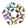
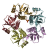
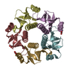
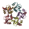
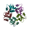
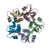
| Entry | Database: PDB / ID: 1qb5 | ||||||
|---|---|---|---|---|---|---|---|
| Title | ESCHERICHIA COLI HEAT LABILE ENTEROTOXIN TYPE IIB B-PENTAMER | ||||||
 Components Components | PROTEIN (HEAT LABILE ENTEROTOXIN TYPE IIB B-PENTAMER) | ||||||
 Keywords Keywords | ENTEROTOXIN | ||||||
| Function / homology |  Function and homology information Function and homology information | ||||||
| Biological species |  | ||||||
| Method |  X-RAY DIFFRACTION / SYNCHROTRON / Resolution: 1.9 Å X-RAY DIFFRACTION / SYNCHROTRON / Resolution: 1.9 Å | ||||||
 Authors Authors | Merritt, E.A. / Hol, W.G.J. | ||||||
 Citation Citation | Journal: To be Published Title: Crystal Structures of the B-Pentamer from E. coli Enterotoxin LT-IIb Authors: Merritt, E.A. / Van den Akker, F. / Connell, T.D. / Holmes, R.K. / Hol, W.G.J. #1: Journal: Structure / Year: 1996Title: Crystal Structure of a New Heat-Labile Enterotoxin, LT-IIb Authors: Van Den Akker, F. / Sarfaty, S. / Twiddy, E.M. / Connell, T.D. / Holmes, R.K. / Hol, W.G.J. #2: Journal: J.Bacteriol. / Year: 1989Title: Cloning, Nucleotide Sequence, and Hybridization Studies of the Type IIb Heat- Labile Enterotoxin Gene of Escherichia coli Authors: Pickett, C.L. / Twiddy, E.M. / Coker, C. / Holmes, R.K. | ||||||
| History |
|
- Structure visualization
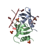
| Structure viewer | Molecule: MolmilJmol/JSmol |
|---|
- Downloads & links
-Download
| PDBx/mmCIF format | 1qb5.cif.gz | 104.6 KB | Display | PDBx/mmCIF format |
|---|---|---|---|---|
| PDB format | pdb1qb5.ent.gz | 82.6 KB | Display | PDB format |
| PDBx/mmJSON format | 1qb5.json.gz | Tree view | PDBx/mmJSON format | |
| Others |  Other downloads Other downloads |
-Validation report
| Arichive directory | https://data.pdbj.org/pub/pdb/validation_reports/qb/1qb5ftp://data.pdbj.org/pub/pdb/validation_reports/qb/1qb5 | HTTPS FTP |
|---|
-Related structure data


-Links
 PDBj
PDBj
- Assembly
| Deposited unit | 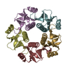
| ||||||||
|---|---|---|---|---|---|---|---|---|---|
| 1 |
| ||||||||
| Unit cell |
|
-Components
| #1: Protein | Mass: 10778.190 Da / Num. of mol.: 5 Source method: isolated from a genetically manipulated source Source: (gene. exp.) #2: Water | ChemComp-HOH / |  Mass: 18.015 Da / Num. of mol.: 188 / Source method: isolated from a natural source / Formula: H2O Mass: 18.015 Da / Num. of mol.: 188 / Source method: isolated from a natural source / Formula: H2OHas protein modification | Y | |
|---|
-Experimental details
-Experiment
| Experiment | Method: X-RAY DIFFRACTION / Number of used crystals: 2 |
|---|
- Sample preparation
| Crystal | Density Matthews: 1.57 Å3/Da / Density % sol: 21.4 % |
|---|---|
| Crystal grow | Method: vapor diffusion, sitting drop / pH: 5.5 / Details: VAPOR DIFFUSION, SITTING DROP, pH 5.5 |
-Data collection
| Diffraction | Mean temperature: 275 K |
|---|---|
| Diffraction source | Source: SYNCHROTRON / Site: SSRL  / Beamline: BL7-1 / Wavelength: 1.08 / Beamline: BL7-1 / Wavelength: 1.08 |
| Detector | Type: MARRESEARCH / Detector: IMAGE PLATE |
| Radiation | Protocol: SINGLE WAVELENGTH / Monochromatic (M) / Laue (L): M / Scattering type: x-ray |
| Radiation wavelength | Wavelength: 1.08 Å / Relative weight: 1 |
| Reflection | Resolution: 1.9→30 Å / Num. all: 36737 / Num. obs: 36737 / % possible obs: 94 % / Rmerge(I) obs: 0.101 / Net I/σ(I): 14 |
| Reflection shell | Resolution: 1.9→1.97 Å / Rmerge(I) obs: 0.335 / Mean I/σ(I) obs: 5.4 / % possible all: 92 |
- Processing
| Software |
| |||||||||||||||||||||||||||||||||
|---|---|---|---|---|---|---|---|---|---|---|---|---|---|---|---|---|---|---|---|---|---|---|---|---|---|---|---|---|---|---|---|---|---|---|
| Refinement | Resolution: 1.9→10 Å / Num. parameters: 1576 / Num. restraintsaints: 3339 / Cross valid method: FREE R / σ(F): 0 Details: ANISOTROPIC SCALING APPLIED BY THE METHOD OF PARKIN, MOEZZI & HOPE, J.APPL.CRYST.28(1995)53-56
| |||||||||||||||||||||||||||||||||
| Solvent computation | Solvent model: MOEWS & KRETSINGER, J.MOL.BIOL.91(1973)201-228 | |||||||||||||||||||||||||||||||||
| Refine analyze | Occupancy sum hydrogen: 3675 / Occupancy sum non hydrogen: 3938 | |||||||||||||||||||||||||||||||||
| Refinement step | Cycle: LAST / Resolution: 1.9→10 Å
| |||||||||||||||||||||||||||||||||
| Refine LS restraints |
|
