- PDB-1q6y: Hypothetical protein YfdW from E. coli bound to Coenzyme A -
+
Open data
ID or keywords:
Loading...
-
Basic information
Entry
Database: PDB / ID: 1q6y
Title
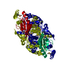
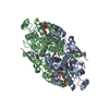
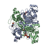
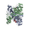
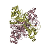
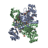
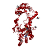
Hypothetical protein YfdW from E. coli bound to Coenzyme A
Components
Hypothetical protein yfdW
Keywords
TRANSFERASE / intertwined dimer / structural genomics / Coenzyme A / PSI / Protein Structure Initiative / New York SGX Research Center for Structural Genomics / NYSGXRC
Function / homology
Function and homology information
formyl-CoA transferase / formyl-CoA transferase activity / oxalate catabolic process / cellular response to acidic pH Similarity search - Function
Formyl-CoA:oxalate CoA-transferase / : / formyl-coa transferase, domain 3 / Crotonobetainyl-coa:carnitine coa-transferase; domain 1 / formyl-coa transferase, domain 3 / CoA-transferase family III / CoA-transferase family III domain 1 superfamily / CoA-transferase family III domain 3 superfamily / CoA-transferase family III / Rossmann fold ...Formyl-CoA:oxalate CoA-transferase / : / formyl-coa transferase, domain 3 / Crotonobetainyl-coa:carnitine coa-transferase; domain 1 / formyl-coa transferase, domain 3 / CoA-transferase family III / CoA-transferase family III domain 1 superfamily / CoA-transferase family III domain 3 superfamily / CoA-transferase family III / Rossmann fold / 2-Layer Sandwich / 3-Layer(aba) Sandwich / Alpha Beta Similarity search - Domain/homology
SEQUENCE RESIDUES SER2 AND LEU3 ARISE FROM A CLONING ARTIFACT THAT RESULTS IN A 2 AMINO ACID ...SEQUENCE RESIDUES SER2 AND LEU3 ARISE FROM A CLONING ARTIFACT THAT RESULTS IN A 2 AMINO ACID INSERTION. RESIDUE NUMBERS AFTER THIS INSERTION ARE EQUAL TO 2 PLUS THE CORRECT RESIDUE NUMBER.
In the structure databanks used in Yorodumi, some data are registered as the other names, "COVID-19 virus" and "2019-nCoV". Here are the details of the virus and the list of structure data.
Jan 31, 2019. EMDB accession codes are about to change! (news from PDBe EMDB page)
EMDB accession codes are about to change! (news from PDBe EMDB page)
The allocation of 4 digits for EMDB accession codes will soon come to an end. Whilst these codes will remain in use, new EMDB accession codes will include an additional digit and will expand incrementally as the available range of codes is exhausted. The current 4-digit format prefixed with “EMD-” (i.e. EMD-XXXX) will advance to a 5-digit format (i.e. EMD-XXXXX), and so on. It is currently estimated that the 4-digit codes will be depleted around Spring 2019, at which point the 5-digit format will come into force.
The EM Navigator/Yorodumi systems omit the EMD- prefix.
Related info.:Q: What is EMD? / ID/Accession-code notation in Yorodumi/EM Navigator
Yorodumi is a browser for structure data from EMDB, PDB, SASBDB, etc.
This page is also the successor to EM Navigator detail page, and also detail information page/front-end page for Omokage search.
The word "yorodu" (or yorozu) is an old Japanese word meaning "ten thousand". "mi" (miru) is to see.
Related info.:EMDB / PDB / SASBDB / Comparison of 3 databanks / Yorodumi Search / Aug 31, 2016. New EM Navigator & Yorodumi / Yorodumi Papers / Jmol/JSmol / Function and homology information / Changes in new EM Navigator and Yorodumi
 Movie
Movie Controller


 Open data
 Basic information
 Components
 Keywords
 Function and homology information
 X-RAY DIFFRACTION /
  Authors
 Citation
 Structure visualization
 Downloads & links
 Other downloads


 PDBj
PDBj


 Assembly

 Mass: 767.534 Da / Num. of mol.: 1 / Source method: obtained synthetically / Formula: C21H36N7O16P3S
 Mass: 118.174 Da / Num. of mol.: 7 / Source method: obtained synthetically / Formula: C6H14O2 / Comment: precipitant*YM
 Mass: 18.015 Da / Num. of mol.: 276 / Source method: isolated from a natural source / Formula: H2O
 Sample preparation
 / Beamline: 31-ID / Wavelength: 0.9793 Å
 Processing