 Movie
Movie Controller


+ Open data

- Basic information
| Entry | Database: PDB / ID: 1q5q | ||||||
|---|---|---|---|---|---|---|---|
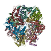
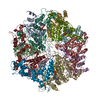
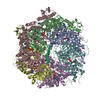
| Title | The Rhodococcus 20S proteasome | ||||||
 Components Components |
| ||||||
 Keywords Keywords | HYDROLASE / proteasome assembly / pro-peptide / inter-subunit contacts / Rhodococcus erythropolis | ||||||
| Function / homology |  Function and homology information Function and homology informationproteasome endopeptidase complex / proteasome core complex, beta-subunit complex / threonine-type endopeptidase activity / proteasome core complex, alpha-subunit complex / proteasomal protein catabolic process / modification-dependent protein catabolic process / endopeptidase activity / cytoplasm Similarity search - Function | ||||||
| Biological species |  Rhodococcus erythropolis (bacteria) Rhodococcus erythropolis (bacteria) | ||||||
| Method |  X-RAY DIFFRACTION / SYNCHROTRON / MOLECULAR REPLACEMENT / Resolution: 2.6 Å X-RAY DIFFRACTION / SYNCHROTRON / MOLECULAR REPLACEMENT / Resolution: 2.6 Å | ||||||
 Authors Authors | Kwon, Y.D. / Nagy, I. / Adams, P.D. / Baumeister, W. / Jap, B.K. | ||||||
 Citation Citation | Journal: J.Mol.Biol. / Year: 2004 Title: Crystal structures of the Rhodococcus proteasome with and without its pro-peptides: implications for the role of the pro-peptide in proteasome assembly. Authors: Kwon, Y.D. / Nagy, I. / Adams, P.D. / Baumeister, W. / Jap, B.K. | ||||||
| History |
|
- Structure visualization
| Structure viewer | Molecule: MolmilJmol/JSmol |
|---|
- Downloads & links
-Download
| PDBx/mmCIF format | 1q5q.cif.gz | 568.7 KB | Display | PDBx/mmCIF format |
|---|---|---|---|---|
| PDB format | pdb1q5q.ent.gz | 472.6 KB | Display | PDB format |
| PDBx/mmJSON format | 1q5q.json.gz | Tree view | PDBx/mmJSON format | |
| Others |  Other downloads Other downloads |
-Validation report
| Arichive directory | https://data.pdbj.org/pub/pdb/validation_reports/q5/1q5qftp://data.pdbj.org/pub/pdb/validation_reports/q5/1q5q | HTTPS FTP |
|---|
-Related structure data
| Related structure data |  1q5rC  1pmaS C: citing same article ( S: Starting model for refinement |
|---|---|
| Similar structure data |


-Links
 PDBj
PDBj


- Assembly
| Deposited unit | 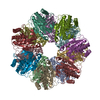
| ||||||||
|---|---|---|---|---|---|---|---|---|---|
| 1 |
| ||||||||
| 2 | 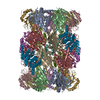
| ||||||||
| Unit cell |
|
-Components
| #1: Protein | Mass: 28346.840 Da / Num. of mol.: 7 Source method: isolated from a genetically manipulated source Source: (gene. exp.) Rhodococcus erythropolis (bacteria) / Strain: BL21 / Gene: PRCA(1) / Plasmid: pT7-7 / Production host: References: UniProt: Q53080, proteasome endopeptidase complex #2: Protein | Mass: 24984.766 Da / Num. of mol.: 7 Source method: isolated from a genetically manipulated source Source: (gene. exp.) Rhodococcus erythropolis (bacteria) / Strain: BL21 / Gene: PRCB(1) / Plasmid: pT7-7 / Production host: #3: Water | ChemComp-HOH / |  Mass: 18.015 Da / Num. of mol.: 167 / Source method: isolated from a natural source / Formula: H2O Mass: 18.015 Da / Num. of mol.: 167 / Source method: isolated from a natural source / Formula: H2O |
|---|
-Experimental details
-Experiment
| Experiment | Method: X-RAY DIFFRACTION / Number of used crystals: 1 |
|---|
- Sample preparation
| Crystal | Density Matthews: 2.72 Å3/Da / Density % sol: 54.71 % | ||||||||||||||||||||||||||||||
|---|---|---|---|---|---|---|---|---|---|---|---|---|---|---|---|---|---|---|---|---|---|---|---|---|---|---|---|---|---|---|---|
| Crystal grow | Temperature: 298 K / Method: vapor diffusion, sitting drop / pH: 5.4 Details: 14% PEG 6000, 50mM sodium citrate, pH 5.4, VAPOR DIFFUSION, SITTING DROP, temperature 298K | ||||||||||||||||||||||||||||||
| Crystal grow | *PLUS pH: 7 / Method: vapor diffusion, sitting drop | ||||||||||||||||||||||||||||||
| Components of the solutions | *PLUS
|
-Data collection
| Diffraction | Mean temperature: 110 K |
|---|---|
| Diffraction source | Source: SYNCHROTRON / Site: ALS  / Beamline: 5.0.1 / Wavelength: 1 Å / Beamline: 5.0.1 / Wavelength: 1 Å |
| Detector | Type: ADSC QUANTUM 4 / Detector: CCD / Date: Nov 3, 2001 |
| Radiation | Protocol: SINGLE WAVELENGTH / Monochromatic (M) / Laue (L): M / Scattering type: x-ray |
| Radiation wavelength | Wavelength: 1 Å / Relative weight: 1 |
| Reflection | Resolution: 2.6→20 Å / Num. obs: 114039 / Observed criterion σ(I): 0 / Biso Wilson estimate: 42.5 Å2 / Rmerge(I) obs: 0.074 |
| Reflection | *PLUS Highest resolution: 2.6 Å / % possible obs: 91.7 % |
- Processing
| Software |
| ||||||||||||||||||||||||||||||||||||
|---|---|---|---|---|---|---|---|---|---|---|---|---|---|---|---|---|---|---|---|---|---|---|---|---|---|---|---|---|---|---|---|---|---|---|---|---|---|
| Refinement | Method to determine structure: MOLECULAR REPLACEMENT Starting model: PDB entry 1PMA Resolution: 2.6→19.99 Å / Rfactor Rfree error: 0.002 / Data cutoff high absF: 183405.38 / Data cutoff high rms absF: 183405.38 / Data cutoff low absF: 0 / Isotropic thermal model: RESTRAINED / Cross valid method: THROUGHOUT / σ(F): 0 / σ(I): 0
| ||||||||||||||||||||||||||||||||||||
| Solvent computation | Solvent model: FLAT MODEL / Bsol: 23.7308 Å2 / ksol: 0.312508 e/Å3 | ||||||||||||||||||||||||||||||||||||
| Displacement parameters | Biso mean: 46.1 Å2
| ||||||||||||||||||||||||||||||||||||
| Refine analyze |
| ||||||||||||||||||||||||||||||||||||
| Refinement step | Cycle: LAST / Resolution: 2.6→19.99 Å
| ||||||||||||||||||||||||||||||||||||
| Refine LS restraints |
| ||||||||||||||||||||||||||||||||||||
| Refine LS restraints NCS | NCS model details: CONSTR | ||||||||||||||||||||||||||||||||||||
| LS refinement shell | Resolution: 2.6→2.76 Å / Rfactor Rfree error: 0.009 / Total num. of bins used: 6
| ||||||||||||||||||||||||||||||||||||
| Xplor file |
| ||||||||||||||||||||||||||||||||||||
| Refinement | *PLUS Highest resolution: 2.6 Å / Lowest resolution: 20 Å / Num. reflection obs: 113987 | ||||||||||||||||||||||||||||||||||||
| Solvent computation | *PLUS | ||||||||||||||||||||||||||||||||||||
| Displacement parameters | *PLUS | ||||||||||||||||||||||||||||||||||||
| Refine LS restraints | *PLUS
|
