 Movie
Movie Controller

[English] 日本語
 Yorodumi
- PDB-1q4s: Crystal structure of Arthrobacter sp. strain SU 4-hydroxybenzoyl ... -
+ Open data

- Basic information
| Entry | Database: PDB / ID: 1q4s | ||||||
|---|---|---|---|---|---|---|---|
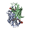
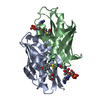
| Title | Crystal structure of Arthrobacter sp. strain SU 4-hydroxybenzoyl CoA thioesterase complexed with CoA and 4-hydroxybenzoic acid | ||||||
 Components Components | Thioesterase | ||||||
 Keywords Keywords | HYDROLASE / thioesterase / hot-dog | ||||||
| Function / homology |  Function and homology information Function and homology information4-hydroxybenzoyl-CoA thioesterase / 4-hydroxybenzoyl-CoA thioesterase activity / 1,4-dihydroxy-2-naphthoyl-CoA thioesterase activity / cytosol Similarity search - Function | ||||||
| Biological species |  Arthrobacter sp. (bacteria) Arthrobacter sp. (bacteria) | ||||||
| Method |  X-RAY DIFFRACTION / MOLECULAR REPLACEMENT / Resolution: 1.95 Å X-RAY DIFFRACTION / MOLECULAR REPLACEMENT / Resolution: 1.95 Å | ||||||
 Authors Authors | Thoden, J.B. / Zhuang, Z. / Dunaway-Mariano, D. / Holden, H.M. | ||||||
 Citation Citation | Journal: J.Biol.Chem. / Year: 2003 Title: The Structure of 4-Hydroxybenzoyl-CoA Thioesterase from Arthrobacter sp. strain SU Authors: Thoden, J.B. / Zhuang, Z. / Dunaway-Mariano, D. / Holden, H.M. | ||||||
| History |
|
- Structure visualization
| Structure viewer | Molecule: MolmilJmol/JSmol |
|---|
- Downloads & links
-Download
| PDBx/mmCIF format | 1q4s.cif.gz | 75.5 KB | Display | PDBx/mmCIF format |
|---|---|---|---|---|
| PDB format | pdb1q4s.ent.gz | 56.2 KB | Display | PDB format |
| PDBx/mmJSON format | 1q4s.json.gz | Tree view | PDBx/mmJSON format | |
| Others |  Other downloads Other downloads |
-Validation report
| Arichive directory | https://data.pdbj.org/pub/pdb/validation_reports/q4/1q4sftp://data.pdbj.org/pub/pdb/validation_reports/q4/1q4s | HTTPS FTP |
|---|
-Related structure data


-Links
 PDBj
PDBj


- Assembly
| Deposited unit | 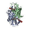
| ||||||||
|---|---|---|---|---|---|---|---|---|---|
| 1 | 
| ||||||||
| Unit cell |
| ||||||||
| Details | homotetramer. The tetramer is generated by expanding the crystallographic independent dimer around the crystallographic 2-fold axis. |
-Components
| #1: Protein | Mass: 16416.428 Da / Num. of mol.: 2 Source method: isolated from a genetically manipulated source Source: (gene. exp.) Arthrobacter sp. (bacteria) / Strain: SU / Gene: FCBC / Plasmid: pET23b / Production host: References: UniProt: Q04416, 4-hydroxybenzoyl-CoA thioesterase #2: Chemical |   Mass: 767.534 Da / Num. of mol.: 2 / Source method: obtained synthetically / Formula: C21H36N7O16P3S Mass: 767.534 Da / Num. of mol.: 2 / Source method: obtained synthetically / Formula: C21H36N7O16P3S#3: Chemical |   Mass: 138.121 Da / Num. of mol.: 2 / Source method: obtained synthetically / Formula: C7H6O3 Mass: 138.121 Da / Num. of mol.: 2 / Source method: obtained synthetically / Formula: C7H6O3#4: Water | ChemComp-HOH / |  Mass: 18.015 Da / Num. of mol.: 241 / Source method: isolated from a natural source / Formula: H2O Mass: 18.015 Da / Num. of mol.: 241 / Source method: isolated from a natural source / Formula: H2O |
|---|
-Experimental details
-Experiment
| Experiment | Method: X-RAY DIFFRACTION / Number of used crystals: 1 |
|---|
- Sample preparation
| Crystal | Density Matthews: 3.38 Å3/Da / Density % sol: 63.59 % | ||||||||||||||||||||||||||||||||||||||||||||||||||||||||
|---|---|---|---|---|---|---|---|---|---|---|---|---|---|---|---|---|---|---|---|---|---|---|---|---|---|---|---|---|---|---|---|---|---|---|---|---|---|---|---|---|---|---|---|---|---|---|---|---|---|---|---|---|---|---|---|---|---|
| Crystal grow | Temperature: 298 K / Method: vapor diffusion, hanging drop / pH: 7.5 Details: PEG-3400, MOPS, LiCl, KCl, 4-hydroxybenzoyl CoA, pH 7.5, VAPOR DIFFUSION, HANGING DROP, temperature 298K | ||||||||||||||||||||||||||||||||||||||||||||||||||||||||
| Crystal grow | *PLUS Temperature: 4 ℃ / Method: vapor diffusion, hanging drop | ||||||||||||||||||||||||||||||||||||||||||||||||||||||||
| Components of the solutions | *PLUS
|
-Data collection
| Diffraction | Mean temperature: 110 K |
|---|---|
| Diffraction source | Source: ROTATING ANODE / Type: RIGAKU RU200 / Wavelength: 1.5418 Å |
| Detector | Type: SIEMENS HI-STAR / Detector: AREA DETECTOR / Date: May 20, 2002 / Details: goebel mirrors |
| Radiation | Monochromator: goebel optics / Protocol: SINGLE WAVELENGTH / Monochromatic (M) / Laue (L): M / Scattering type: x-ray |
| Radiation wavelength | Wavelength: 1.5418 Å / Relative weight: 1 |
| Reflection | Resolution: 1.95→30 Å / Num. all: 30625 / Num. obs: 30625 / % possible obs: 94.1 % / Observed criterion σ(F): 0 / Observed criterion σ(I): 0 / Redundancy: 3.6 % / Rsym value: 0.051 / Net I/σ(I): 24.6 |
| Reflection shell | Resolution: 1.95→2.04 Å / Redundancy: 1.9 % / Mean I/σ(I) obs: 4.1 / Num. unique all: 3403 / Rsym value: 0.22 / % possible all: 83.4 |
| Reflection | *PLUS Rmerge(I) obs: 0.051 |
| Reflection shell | *PLUS % possible obs: 83.4 % / Num. unique obs: 3403 / Rmerge(I) obs: 0.22 |
- Processing
| Software |
| |||||||||||||||||||||||||
|---|---|---|---|---|---|---|---|---|---|---|---|---|---|---|---|---|---|---|---|---|---|---|---|---|---|---|
| Refinement | Method to determine structure: MOLECULAR REPLACEMENT / Resolution: 1.95→30 Å / Cross valid method: THROUGHOUT / σ(F): 0 / σ(I): 0 / Stereochemistry target values: Engh & Huber
| |||||||||||||||||||||||||
| Refinement step | Cycle: LAST / Resolution: 1.95→30 Å
| |||||||||||||||||||||||||
| Refine LS restraints |
| |||||||||||||||||||||||||
| Refinement | *PLUS Num. reflection obs: 27534 | |||||||||||||||||||||||||
| Solvent computation | *PLUS | |||||||||||||||||||||||||
| Displacement parameters | *PLUS | |||||||||||||||||||||||||
| Refine LS restraints | *PLUS
|
