 Movie
Movie Controller

[English] 日本語
 Yorodumi
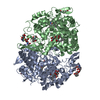
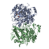
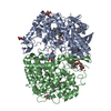
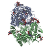
- PDB-1q4g: 2.0 Angstrom Crystal Structure of Ovine Prostaglandin H2 Synthase... -
+ Open data

- Basic information
| Entry | Database: PDB / ID: 1q4g | |||||||||
|---|---|---|---|---|---|---|---|---|---|---|
| Title | 2.0 Angstrom Crystal Structure of Ovine Prostaglandin H2 Synthase-1, in complex with alpha-methyl-4-biphenylacetic acid | |||||||||
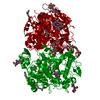
 Components Components | Prostaglandin G/H synthase 1 | |||||||||
 Keywords Keywords | OXIDOREDUCTASE / cyclooxygenase / non-steroidal anti-inflammatory drug / peroxidase / prostaglandin synthase / EGF-like domain / membrane binding domain | |||||||||
| Function / homology |  Function and homology information Function and homology informationprostaglandin-endoperoxide synthase / prostaglandin-endoperoxide synthase activity / cyclooxygenase pathway / oxidoreductase activity, acting on single donors with incorporation of molecular oxygen, incorporation of two atoms of oxygen / prostaglandin biosynthetic process / peroxidase activity / regulation of blood pressure / response to oxidative stress / neuron projection / heme binding ...prostaglandin-endoperoxide synthase / prostaglandin-endoperoxide synthase activity / cyclooxygenase pathway / oxidoreductase activity, acting on single donors with incorporation of molecular oxygen, incorporation of two atoms of oxygen / prostaglandin biosynthetic process / peroxidase activity / regulation of blood pressure / response to oxidative stress / neuron projection / heme binding / endoplasmic reticulum membrane / protein homodimerization activity / metal ion binding / cytoplasm Similarity search - Function | |||||||||
| Biological species |  | |||||||||
| Method |  X-RAY DIFFRACTION / SYNCHROTRON / MOLECULAR REPLACEMENT / Resolution: 2 Å X-RAY DIFFRACTION / SYNCHROTRON / MOLECULAR REPLACEMENT / Resolution: 2 Å | |||||||||
 Authors Authors | Gupta, K. / Selinksy, B.S. / Kaub, C.J. / Katz, A.K. / Loll, P.J. | |||||||||
 Citation Citation | Journal: J.Mol.Biol. / Year: 2004 Title: The 2.0A Resolution Crystal Structure of Prostaglandin H(2) Synthase-1: Structural Insights into an Unusual Peroxidase Authors: Gupta, K. / Selinsky, B.S. / Kaub, C.J. / Katz, A.K. / Loll, P.J. | |||||||||
| History |
|
- Structure visualization
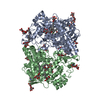
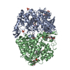
| Structure viewer | Molecule: MolmilJmol/JSmol |
|---|
- Downloads & links
-Download
| PDBx/mmCIF format | 1q4g.cif.gz | 265.8 KB | Display | PDBx/mmCIF format |
|---|---|---|---|---|
| PDB format | pdb1q4g.ent.gz | 213.8 KB | Display | PDB format |
| PDBx/mmJSON format | 1q4g.json.gz | Tree view | PDBx/mmJSON format | |
| Others |  Other downloads Other downloads |
-Validation report
| Arichive directory | https://data.pdbj.org/pub/pdb/validation_reports/q4/1q4gftp://data.pdbj.org/pub/pdb/validation_reports/q4/1q4g | HTTPS FTP |
|---|
-Related structure data
| Related structure data |  1eqhS S: Starting model for refinement |
|---|---|
| Similar structure data |


-Links
 PDBj
PDBj


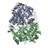
- Assembly
| Deposited unit | 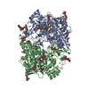
| ||||||||
|---|---|---|---|---|---|---|---|---|---|
| 1 |
| ||||||||
| Unit cell |
|
-Components
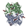
-Protein , 1 types, 2 molecules AB
| #1: Protein | Mass: 63724.141 Da / Num. of mol.: 2 / Source method: isolated from a natural source / Source: (natural) References: GenBank: 165844, UniProt: P05979*PLUS, prostaglandin-endoperoxide synthase |
|---|
-Sugars , 5 types, 14 molecules 
| #2: Polysaccharide | Source method: isolated from a genetically manipulated source #3: Polysaccharide | Source method: isolated from a genetically manipulated source #4: Polysaccharide | alpha-D-mannopyranose-(1-6)-alpha-D-mannopyranose-(1-6)-beta-D-mannopyranose-(1-4)-2-acetamido-2- ...alpha-D-mannopyranose-(1-6)-alpha-D-mannopyranose-(1-6)-beta-D-mannopyranose-(1-4)-2-acetamido-2-deoxy-alpha-D-glucopyranose-(1-4)-2-acetamido-2-deoxy-beta-D-glucopyranose | Source method: isolated from a genetically manipulated source #5: Polysaccharide | beta-D-mannopyranose-(1-6)-beta-D-mannopyranose-(1-6)-beta-D-mannopyranose-(1-4)-2-acetamido-2- ...beta-D-mannopyranose-(1-6)-beta-D-mannopyranose-(1-6)-beta-D-mannopyranose-(1-4)-2-acetamido-2-deoxy-alpha-D-glucopyranose-(1-4)-2-acetamido-2-deoxy-beta-D-glucopyranose | Source method: isolated from a genetically manipulated source #6: Sugar | ChemComp-BOG /  Type: D-saccharide / Mass: 292.369 Da / Num. of mol.: 8 Type: D-saccharide / Mass: 292.369 Da / Num. of mol.: 8Source method: isolated from a genetically manipulated source Formula: C14H28O6 / Comment: detergent*YM |
|---|
-Non-polymers , 4 types, 663 molecules 


| #7: Chemical |  Mass: 226.270 Da / Num. of mol.: 2 / Source method: obtained synthetically / Formula: C15H14O2 Mass: 226.270 Da / Num. of mol.: 2 / Source method: obtained synthetically / Formula: C15H14O2#8: Chemical |  Mass: 616.487 Da / Num. of mol.: 2 / Source method: obtained synthetically / Formula: C34H32FeN4O4 Mass: 616.487 Da / Num. of mol.: 2 / Source method: obtained synthetically / Formula: C34H32FeN4O4#9: Chemical | ChemComp-GOL /  Mass: 92.094 Da / Num. of mol.: 5 / Source method: obtained synthetically / Formula: C3H8O3 Mass: 92.094 Da / Num. of mol.: 5 / Source method: obtained synthetically / Formula: C3H8O3#10: Water | ChemComp-HOH / | Mass: 18.015 Da / Num. of mol.: 654 / Source method: isolated from a natural source / Formula: H2O |
|---|
-Details
| Has protein modification | Y |
|---|
-Experimental details
-Experiment
| Experiment | Method: X-RAY DIFFRACTION / Number of used crystals: 1 |
|---|
- Sample preparation
| Crystal | Density Matthews: 4.39 Å3/Da / Density % sol: 71.95 % | ||||||||||||||||||||||||||||||
|---|---|---|---|---|---|---|---|---|---|---|---|---|---|---|---|---|---|---|---|---|---|---|---|---|---|---|---|---|---|---|---|
| Crystal grow | Temperature: 291 K / Method: vapor diffusion, hanging drop / pH: 6.7 Details: 20 mM sodium phosphate pH 6.7, 100-200mM NACL, 0.6% BOG, 1mM NSAID against reservoir of 4-8% PEG-4000, VAPOR DIFFUSION, HANGING DROP, temperature 291K | ||||||||||||||||||||||||||||||
| Crystal grow | *PLUS Temperature: 18 ℃ / Method: vapor diffusion, hanging drop | ||||||||||||||||||||||||||||||
| Components of the solutions | *PLUS
|
-Data collection
| Diffraction | Mean temperature: 180 K |
|---|---|
| Diffraction source | Source: SYNCHROTRON / Site: NSLS  / Beamline: X25 / Wavelength: 1.099 Å / Beamline: X25 / Wavelength: 1.099 Å |
| Detector | Type: ADSC QUANTUM 4 / Detector: CCD / Date: Sep 1, 2002 |
| Radiation | Protocol: SINGLE WAVELENGTH / Monochromatic (M) / Laue (L): M / Scattering type: x-ray |
| Radiation wavelength | Wavelength: 1.099 Å / Relative weight: 1 |
| Reflection | Resolution: 2→50 Å / Num. all: 144508 / Num. obs: 144508 / % possible obs: 94.9 % / Observed criterion σ(F): 0 / Observed criterion σ(I): 2 / Redundancy: 6.63 % / Biso Wilson estimate: 31.3 Å2 / Rmerge(I) obs: 0.103 / Net I/σ(I): 7.8 |
| Reflection shell | Resolution: 2→2.07 Å / Redundancy: 4.3 % / Rmerge(I) obs: 0.52 / Mean I/σ(I) obs: 2.2 / Num. unique all: 9630 / % possible all: 71 |
| Reflection | *PLUS Num. obs: 141441 / Num. measured all: 966916 |
| Reflection shell | *PLUS Highest resolution: 2 Å / Lowest resolution: 2.13 Å / % possible obs: 75.2 % |
- Processing
| Software |
| ||||||||||||||||||||||||||||||||||||
|---|---|---|---|---|---|---|---|---|---|---|---|---|---|---|---|---|---|---|---|---|---|---|---|---|---|---|---|---|---|---|---|---|---|---|---|---|---|
| Refinement | Method to determine structure: MOLECULAR REPLACEMENT Starting model: 1EQH Resolution: 2→43.68 Å / Rfactor Rfree error: 0.002 / Data cutoff high absF: 2580366.56 / Data cutoff low absF: 0 / Isotropic thermal model: RESTRAINED / Cross valid method: THROUGHOUT / σ(F): 0 / σ(I): 2 / Stereochemistry target values: Engh & Huber
| ||||||||||||||||||||||||||||||||||||
| Solvent computation | Solvent model: FLAT MODEL / Bsol: 46.9365 Å2 / ksol: 0.341906 e/Å3 | ||||||||||||||||||||||||||||||||||||
| Displacement parameters | Biso mean: 44.1 Å2
| ||||||||||||||||||||||||||||||||||||
| Refine analyze |
| ||||||||||||||||||||||||||||||||||||
| Refinement step | Cycle: LAST / Resolution: 2→43.68 Å
| ||||||||||||||||||||||||||||||||||||
| Refine LS restraints |
| ||||||||||||||||||||||||||||||||||||
| LS refinement shell | Resolution: 2→2.13 Å / Rfactor Rfree error: 0.009 / Total num. of bins used: 6
| ||||||||||||||||||||||||||||||||||||
| Xplor file |
| ||||||||||||||||||||||||||||||||||||
| Refinement | *PLUS Highest resolution: 2 Å / Lowest resolution: 50 Å | ||||||||||||||||||||||||||||||||||||
| Solvent computation | *PLUS | ||||||||||||||||||||||||||||||||||||
| Displacement parameters | *PLUS | ||||||||||||||||||||||||||||||||||||
| Refine LS restraints | *PLUS
|
