 Movie
Movie Controller

[English] 日本語
 Yorodumi
- PDB-1q0q: Crystal structure of DXR in complex with the substrate 1-deoxy-D-... -
+ Open data

- Basic information
| Entry | Database: PDB / ID: 1q0q | ||||||
|---|---|---|---|---|---|---|---|
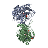
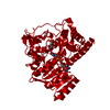
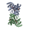
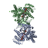
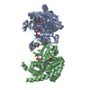
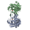
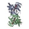
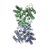
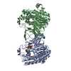
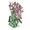
| Title | Crystal structure of DXR in complex with the substrate 1-deoxy-D-xylulose-5-phosphate | ||||||
 Components Components | 1-deoxy-D-xylulose 5-phosphate reductoisomerase | ||||||
 Keywords Keywords | OXIDOREDUCTASE | ||||||
| Function / homology |  Function and homology information Function and homology informationDxr protein complex / isopentenyl diphosphate biosynthetic process, methylerythritol 4-phosphate pathway involved in terpenoid biosynthetic process / 1-deoxy-D-xylulose-5-phosphate reductoisomerase / 1-deoxy-D-xylulose-5-phosphate reductoisomerase activity / isopentenyl diphosphate biosynthetic process, methylerythritol 4-phosphate pathway / NADPH binding / manganese ion binding / identical protein binding Similarity search - Function | ||||||
| Biological species |  | ||||||
| Method |  X-RAY DIFFRACTION / SYNCHROTRON / MOLECULAR REPLACEMENT / Resolution: 1.9 Å X-RAY DIFFRACTION / SYNCHROTRON / MOLECULAR REPLACEMENT / Resolution: 1.9 Å | ||||||
 Authors Authors | Mac Sweeney, A. / Lange, R. / D'Arcy, A. / Douangamath, A. / Surivet, J.-P. / Oefner, C. | ||||||
 Citation Citation | Journal: J.Mol.Biol. / Year: 2005 Title: The crystal structure of E.coli 1-deoxy-D-xylulose-5-phosphate reductoisomerase in a ternary complex with the antimalarial compound fosmidomycin and NADPH reveals a tight-binding closed enzyme conformation. Authors: Mac Sweeney, A. / Lange, R. / Fernandes, R.P. / Schulz, H. / Dale, G.E. / Douangamath, A. / Proteau, P.J. / Oefner, C. | ||||||
| History |
|
- Structure visualization
| Structure viewer | Molecule: MolmilJmol/JSmol |
|---|
- Downloads & links
-Download
| PDBx/mmCIF format | 1q0q.cif.gz | 179.7 KB | Display | PDBx/mmCIF format |
|---|---|---|---|---|
| PDB format | pdb1q0q.ent.gz | 141.1 KB | Display | PDB format |
| PDBx/mmJSON format | 1q0q.json.gz | Tree view | PDBx/mmJSON format | |
| Others |  Other downloads Other downloads |
-Validation report
| Summary document | 1q0q_validation.pdf.gz | 1.1 MB | Display | wwPDB validaton report |
|---|---|---|---|---|
| Full document | 1q0q_full_validation.pdf.gz | 1.1 MB | Display | |
| Data in XML | 1q0q_validation.xml.gz | 37.4 KB | Display | |
| Data in CIF | 1q0q_validation.cif.gz | 55.4 KB | Display | |
| Arichive directory | https://data.pdbj.org/pub/pdb/validation_reports/q0/1q0qftp://data.pdbj.org/pub/pdb/validation_reports/q0/1q0q | HTTPS FTP |
-Related structure data
| Related structure data | 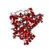 1q0hSC  1q0lC S: Starting model for refinement C: citing same article ( |
|---|---|
| Similar structure data |
-Links
 PDBj
PDBj

- Assembly
| Deposited unit | 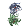
| ||||||||
|---|---|---|---|---|---|---|---|---|---|
| 1 |
| ||||||||
| Unit cell |
| ||||||||
| Details | Biological homodimer contained in the asymmetric unit |
-Components
| #1: Protein | Mass: 44404.879 Da / Num. of mol.: 2 Source method: isolated from a genetically manipulated source Source: (gene. exp.) References: UniProt: P45568, 1-deoxy-D-xylulose-5-phosphate reductoisomerase #2: Chemical |   Mass: 214.110 Da / Num. of mol.: 2 Mass: 214.110 Da / Num. of mol.: 2Source method: isolated from a genetically manipulated source Formula: C5H11O7P #3: Chemical |   Mass: 745.421 Da / Num. of mol.: 2 / Source method: obtained synthetically / Formula: C21H30N7O17P3 Mass: 745.421 Da / Num. of mol.: 2 / Source method: obtained synthetically / Formula: C21H30N7O17P3#4: Water | ChemComp-HOH / |  Mass: 18.015 Da / Num. of mol.: 639 / Source method: isolated from a natural source / Formula: H2O Mass: 18.015 Da / Num. of mol.: 639 / Source method: isolated from a natural source / Formula: H2O |
|---|
-Experimental details
-Experiment
| Experiment | Method: X-RAY DIFFRACTION / Number of used crystals: 1 |
|---|
- Sample preparation
| Crystal | Density Matthews: 2.38 Å3/Da / Density % sol: 48.31 % |
|---|---|
| Crystal grow | Temperature: 298 K / Method: microbatch under oil / pH: 6.5 Details: 25% PEG3350, 200mM Li2SO4, 100mM Bis-Tris pH 6.5, Microbatch under oil, temperature 298K |
-Data collection
| Diffraction | Mean temperature: 100 K |
|---|---|
| Diffraction source | Source: SYNCHROTRON / Site: SLS  / Beamline: X06SA / Wavelength: 0.97818 Å / Beamline: X06SA / Wavelength: 0.97818 Å |
| Detector | Type: MARRESEARCH / Detector: CCD / Date: Apr 10, 2003 |
| Radiation | Protocol: SINGLE WAVELENGTH / Monochromatic (M) / Laue (L): M / Scattering type: x-ray |
| Radiation wavelength | Wavelength: 0.97818 Å / Relative weight: 1 |
| Reflection | Resolution: 1.9→20 Å / Num. all: 62896 / Num. obs: 61149 / % possible obs: 94.2 % / Observed criterion σ(F): 6 / Observed criterion σ(I): 6 / Redundancy: 4.8 % / Rmerge(I) obs: 0.095 / Net I/σ(I): 12.6 |
| Reflection shell | Resolution: 1.9→2.02 Å / Rmerge(I) obs: 0.359 / Mean I/σ(I) obs: 2.73 / % possible all: 95.3 |
- Processing
| Software |
| |||||||||||||||||||||||||
|---|---|---|---|---|---|---|---|---|---|---|---|---|---|---|---|---|---|---|---|---|---|---|---|---|---|---|
| Refinement | Method to determine structure: MOLECULAR REPLACEMENT Starting model: PDB entry 1Q0H Resolution: 1.9→20 Å / Isotropic thermal model: Isotropic / Cross valid method: THROUGHOUT / σ(F): 6 / Stereochemistry target values: Engh & Huber
| |||||||||||||||||||||||||
| Refinement step | Cycle: LAST / Resolution: 1.9→20 Å
| |||||||||||||||||||||||||
| Refine LS restraints |
|
