- PDB-1pzq: Structure of fused docking domains from the erythromycin polyketi... -
+
Open data
ID or keywords:
Loading...
-
Basic information
Entry
Database: PDB / ID: 1pzq
Title
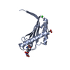
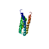
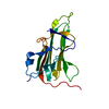
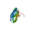
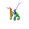
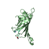
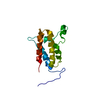
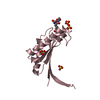
Structure of fused docking domains from the erythromycin polyketide synthase (DEBS), a model for the interaction between DEBS 2 and DEBS 3: The A domain
Erythronolidesynthase / ORF 2 / 6-deoxyerythronolide B synthase II / DEBS 2
Mass: 6428.874 Da / Num. of mol.: 2 / Fragment: C-terminal fragment / Mutation: L1G, F2S Source method: isolated from a genetically manipulated source Source: (gene. exp.) Saccharopolyspora erythraea (bacteria) / Gene: ERYA / Plasmid: pGEX4T-3 / Production host: Escherichia coli (E. coli) / Strain (production host): BL21-CODONPLUS / Keywords: Insertion of GS at N-terminus References: UniProt: Q03132, 6-deoxyerythronolide-B synthase
-
Experimental details
-
Experiment
Experiment
Method: SOLUTION NMR
NMR experiment
Conditions-ID
Experiment-ID
Solution-ID
Type
1
1
1
3D 15N-separated NOESY
2
2
1
3D 13C-separated NOESY
2
3
2
3D 13C 15N X-filtered 13C-separated NOESY
NMR details
Text: The structure was determined using triple-resonance NMR spectroscopy. Intermolecular contacts were obtained from an X-filtered NOESY experiment on a mixed-labeled sample.
Ionic strength: 100mM phosphate buffer NA / pH: 6.5 / Pressure: ambient / Temperature: 298 K
Crystal grow
*PLUS
Method: other / Details: NMR
-
NMR measurement
Radiation
Protocol: SINGLE WAVELENGTH / Monochromatic (M) / Laue (L): M
Radiation wavelength
Relative weight: 1
NMR spectrometer
Type
Manufacturer
Model
Field strength (MHz)
Spectrometer-ID
Bruker DRX
Bruker
DRX
500
1
Bruker DRX
Bruker
DRX
800
2
-
Processing
NMR software
Name
Version
Developer
Classification
ANSIG
3.3
Kraulis
dataanalysis
CNS
1
Brunger
structuresolution
CNS
1
Brunger
refinement
Refinement
Method: simulated annealing / Software ordinal: 1 Details: the structures are based on a total of 1811 restraints: 1709 NOE-derived distance constraints, 60 dihedral angle restraints, 42 distance restraints from hydrogen bonds.
NMR representative
Selection criteria: closest to the average
NMR ensemble
Conformer selection criteria: structures with the lowest energy Conformers calculated total number: 40 / Conformers submitted total number: 8
+
About Yorodumi
-
News
-
Feb 9, 2022. New format data for meta-information of EMDB entries
New format data for meta-information of EMDB entries
Version 3 of the EMDB header file is now the official format.
The previous official version 1.9 will be removed from the archive.
In the structure databanks used in Yorodumi, some data are registered as the other names, "COVID-19 virus" and "2019-nCoV". Here are the details of the virus and the list of structure data.
Jan 31, 2019. EMDB accession codes are about to change! (news from PDBe EMDB page)
EMDB accession codes are about to change! (news from PDBe EMDB page)
The allocation of 4 digits for EMDB accession codes will soon come to an end. Whilst these codes will remain in use, new EMDB accession codes will include an additional digit and will expand incrementally as the available range of codes is exhausted. The current 4-digit format prefixed with “EMD-” (i.e. EMD-XXXX) will advance to a 5-digit format (i.e. EMD-XXXXX), and so on. It is currently estimated that the 4-digit codes will be depleted around Spring 2019, at which point the 5-digit format will come into force.
The EM Navigator/Yorodumi systems omit the EMD- prefix.
Related info.:Q: What is EMD? / ID/Accession-code notation in Yorodumi/EM Navigator
Yorodumi is a browser for structure data from EMDB, PDB, SASBDB, etc.
This page is also the successor to EM Navigator detail page, and also detail information page/front-end page for Omokage search.
The word "yorodu" (or yorozu) is an old Japanese word meaning "ten thousand". "mi" (miru) is to see.
Related info.:EMDB / PDB / SASBDB / Comparison of 3 databanks / Yorodumi Search / Aug 31, 2016. New EM Navigator & Yorodumi / Yorodumi Papers / Jmol/JSmol / Function and homology information / Changes in new EM Navigator and Yorodumi
 Movie
Movie Controller

 Yorodumi
 Open data
 Basic information
 Components
 Keywords
 Function and homology information
 Saccharopolyspora erythraea (bacteria)
 Authors
 Citation
 Structure visualization
 Downloads & links
 Other downloads
 PDBj
PDBj


 Assembly
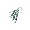
 Sample preparation
 Processing