 Movie
Movie Controller

[English] 日本語
 Yorodumi
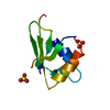
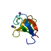
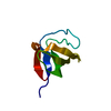
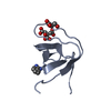
- PDB-1pwt: THERMODYNAMIC ANALYSIS OF ALPHA-SPECTRIN SH3 AND TWO OF ITS CIRCU... -
+ Open data

- Basic information
| Entry | Database: PDB / ID: 1pwt | ||||||
|---|---|---|---|---|---|---|---|
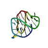
| Title | THERMODYNAMIC ANALYSIS OF ALPHA-SPECTRIN SH3 AND TWO OF ITS CIRCULAR PERMUTANTS WITH DIFFERENT LOOP LENGTHS: DISCERNING THE REASONS FOR RAPID FOLDING IN PROTEINS | ||||||
 Components Components | ALPHA SPECTRIN | ||||||
 Keywords Keywords | CIRCULAR PERMUTANT / SH3 DOMAIN / CYTOSKELETON | ||||||
| Function / homology |  Function and homology information Function and homology informationcostamere / actin filament capping / cortical actin cytoskeleton / cell projection / actin filament binding / cell junction / actin cytoskeleton organization / calmodulin binding / calcium ion binding / plasma membrane Similarity search - Function | ||||||
| Biological species |  | ||||||
| Method |  X-RAY DIFFRACTION / MOLECULAR REPLACEMENT / Resolution: 1.77 Å X-RAY DIFFRACTION / MOLECULAR REPLACEMENT / Resolution: 1.77 Å | ||||||
 Authors Authors | Martinez, J.C. / Viguera, A.R. / Berisio, R. / Wilmanns, M. / Mateo, P.L. / Filmonov, V.V. / Serrano, L. | ||||||
 Citation Citation | Journal: Biochemistry / Year: 1999 Title: Thermodynamic analysis of alpha-spectrin SH3 and two of its circular permutants with different loop lengths: discerning the reasons for rapid folding in proteins. Authors: Martinez, J.C. / Viguera, A.R. / Berisio, R. / Wilmanns, M. / Mateo, P.L. / Filimonov, V.V. / Serrano, L. | ||||||
| History |
|
- Structure visualization
| Structure viewer | Molecule: MolmilJmol/JSmol |
|---|
- Downloads & links
-Download
| PDBx/mmCIF format | 1pwt.cif.gz | 27.3 KB | Display | PDBx/mmCIF format |
|---|---|---|---|---|
| PDB format | pdb1pwt.ent.gz | 16 KB | Display | PDB format |
| PDBx/mmJSON format | 1pwt.json.gz | Tree view | PDBx/mmJSON format | |
| Others |  Other downloads Other downloads |
-Validation report
| Arichive directory | https://data.pdbj.org/pub/pdb/validation_reports/pw/1pwtftp://data.pdbj.org/pub/pdb/validation_reports/pw/1pwt | HTTPS FTP |
|---|
-Related structure data
| Related structure data | 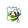 1shgS S: Starting model for refinement |
|---|---|
| Similar structure data |


-Links
 PDBj
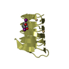
PDBj- Assembly
| Deposited unit | 
| ||||||||
|---|---|---|---|---|---|---|---|---|---|
| 1 |
| ||||||||
| Unit cell |
|
-Components
| #1: Protein | Mass: 7042.095 Da / Num. of mol.: 1 / Fragment: SH3 DOMAIN / Mutation: MGTG INSTEAD OF MDETG AT THE N-TERMINUS / Source method: isolated from a natural source / Source: (natural) |
|---|---|
| #2: Water | ChemComp-HOH /  Mass: 18.015 Da / Num. of mol.: 88 / Source method: isolated from a natural source / Formula: H2O Mass: 18.015 Da / Num. of mol.: 88 / Source method: isolated from a natural source / Formula: H2O |
-Experimental details
-Experiment
| Experiment | Method: X-RAY DIFFRACTION / Number of used crystals: 1 |
|---|
- Sample preparation
| Crystal | Density Matthews: 2.5 Å3/Da / Density % sol: 50 % | ||||||||||||||||||||||||||||||||||||||||
|---|---|---|---|---|---|---|---|---|---|---|---|---|---|---|---|---|---|---|---|---|---|---|---|---|---|---|---|---|---|---|---|---|---|---|---|---|---|---|---|---|---|
| Crystal grow | pH: 4 / Details: pH 4 | ||||||||||||||||||||||||||||||||||||||||
| Crystal | *PLUS | ||||||||||||||||||||||||||||||||||||||||
| Crystal grow | *PLUS Method: vapor diffusion, hanging drop | ||||||||||||||||||||||||||||||||||||||||
| Components of the solutions | *PLUS
|
-Data collection
| Diffraction | Mean temperature: 287 K |
|---|---|
| Diffraction source | Type: OTHER / Wavelength: 1.5418 |
| Detector | Type: MAR scanner 180 mm plate / Detector: IMAGE PLATE |
| Radiation | Monochromatic (M) / Laue (L): M / Scattering type: x-ray |
| Radiation wavelength | Wavelength: 1.5418 Å / Relative weight: 1 |
| Reflection | Resolution: 1.77→15 Å / Num. obs: 7163 / % possible obs: 99.8 % / Redundancy: 6.1 % / Biso Wilson estimate: 18.9 Å2 / Rmerge(I) obs: 0.05 / Net I/σ(I): 25.1 |
| Reflection shell | Resolution: 1.77→1.8 Å / Rmerge(I) obs: 0.2 / Mean I/σ(I) obs: 5.9 / % possible all: 96.8 |
| Reflection | *PLUS Num. measured all: 43628 |
| Reflection shell | *PLUS % possible obs: 96.8 % |
- Processing
| Software |
| ||||||||||||||||||||||||||||||||||||||||||||||||||||||||||||||||||||||||||||||||||||
|---|---|---|---|---|---|---|---|---|---|---|---|---|---|---|---|---|---|---|---|---|---|---|---|---|---|---|---|---|---|---|---|---|---|---|---|---|---|---|---|---|---|---|---|---|---|---|---|---|---|---|---|---|---|---|---|---|---|---|---|---|---|---|---|---|---|---|---|---|---|---|---|---|---|---|---|---|---|---|---|---|---|---|---|---|---|
| Refinement | Method to determine structure: MOLECULAR REPLACEMENT Starting model: 1SHG Resolution: 1.77→14.5 Å / Cross valid method: THROUGHOUT / σ(F): 0 Details: THE N-TERMINUS HAD PROBABLY AN ALTERNATIVE CONFORMATION, BUT THE DENSITY WAS NOT CLEAR ENOUGH FOR MODELLING.
| ||||||||||||||||||||||||||||||||||||||||||||||||||||||||||||||||||||||||||||||||||||
| Displacement parameters | Biso mean: 22.9 Å2 | ||||||||||||||||||||||||||||||||||||||||||||||||||||||||||||||||||||||||||||||||||||
| Refinement step | Cycle: LAST / Resolution: 1.77→14.5 Å
| ||||||||||||||||||||||||||||||||||||||||||||||||||||||||||||||||||||||||||||||||||||
| Refine LS restraints |
|
