 Movie
Movie Controller

[English] 日本語
 Yorodumi
- PDB-1pwh: Rat Liver L-Serine Dehydratase- Complex with PYRIDOXYL-(O-METHYL-... -
+ Open data

- Basic information
| Entry | Database: PDB / ID: 1pwh | ||||||
|---|---|---|---|---|---|---|---|
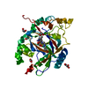
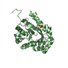
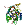
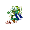
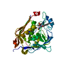
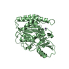
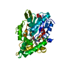
| Title | Rat Liver L-Serine Dehydratase- Complex with PYRIDOXYL-(O-METHYL-SERINE)-5-MONOPHOSPHATE | ||||||
 Components Components | L-serine dehydratase | ||||||
 Keywords Keywords | LYASE / Rat Liver / L-Serine Dehydratase / Complex | ||||||
| Function / homology |  Function and homology information Function and homology informationThreonine catabolism / Serine metabolism / threonine ammonia-lyase / threonine deaminase activity / L-serine ammonia-lyase / L-serine ammonia-lyase activity / response to cobalamin / L-threonine catabolic process / L-serine catabolic process / pyruvate biosynthetic process ...Threonine catabolism / Serine metabolism / threonine ammonia-lyase / threonine deaminase activity / L-serine ammonia-lyase / L-serine ammonia-lyase activity / response to cobalamin / L-threonine catabolic process / L-serine catabolic process / pyruvate biosynthetic process / response to amino acid / gluconeogenesis / lipid metabolic process / response to nutrient levels / pyridoxal phosphate binding / protein-containing complex assembly / protein homodimerization activity / cytoplasm Similarity search - Function | ||||||
| Biological species |  | ||||||
| Method |  X-RAY DIFFRACTION / MOLECULAR REPLACEMENT / Resolution: 2.6 Å X-RAY DIFFRACTION / MOLECULAR REPLACEMENT / Resolution: 2.6 Å | ||||||
 Authors Authors | Yamada, T. / Komoto, J. / Takata, Y. / Ogawa, H. / Takusagawa, F. | ||||||
 Citation Citation | Journal: Biochemistry / Year: 2003 Title: Crystal structure of serine dehydratase from rat liver. Authors: Yamada, T. / Komoto, J. / Takata, Y. / Ogawa, H. / Pitot, H.C. / Takusagawa, F. | ||||||
| History |
| ||||||
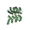
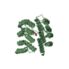
| Remark 300 | BIOMOLECULE: 1, 2, 3, 4 THIS ENTRY CONTAINS THE CRYSTALLOGRAPHIC ASYMMETRIC UNIT WHICH CONSISTS OF ...BIOMOLECULE: 1, 2, 3, 4 THIS ENTRY CONTAINS THE CRYSTALLOGRAPHIC ASYMMETRIC UNIT WHICH CONSISTS OF 4 CHAIN(S). SEE REMARK 350 FOR INFORMATION ON GENERATING THE BIOLOGICAL MOLECULE(S). THE AUTHORS STATE THAT CHAINS AB, CD FORM HOMODIMERS IN TERMS OF STRUCTURE. HOWEVER, SINCE THE ACTIVE SITE IN ONE SUBUNIT IS INDEPENDENT FROM THE OTHER SUBUNIT, THE ENZYMATIC REACTION PROCEEDS MONOMERICALLY. |
- Structure visualization
| Structure viewer | Molecule: MolmilJmol/JSmol |
|---|
- Downloads & links
-Download
| PDBx/mmCIF format | 1pwh.cif.gz | 247.9 KB | Display | PDBx/mmCIF format |
|---|---|---|---|---|
| PDB format | pdb1pwh.ent.gz | 200.8 KB | Display | PDB format |
| PDBx/mmJSON format | 1pwh.json.gz | Tree view | PDBx/mmJSON format | |
| Others |  Other downloads Other downloads |
-Validation report
| Arichive directory | https://data.pdbj.org/pub/pdb/validation_reports/pw/1pwhftp://data.pdbj.org/pub/pdb/validation_reports/pw/1pwh | HTTPS FTP |
|---|
-Related structure data
| Related structure data |  1pweSC S: Starting model for refinement C: citing same article ( |
|---|---|
| Similar structure data |

-Links
 PDBj
PDBj
- Assembly
| Deposited unit | 
| ||||||||
|---|---|---|---|---|---|---|---|---|---|
| 1 | 
| ||||||||
| 2 | 
| ||||||||
| 3 | 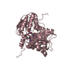
| ||||||||
| 4 | 
| ||||||||
| Unit cell |
|
-Components
| #1: Protein | Mass: 34506.129 Da / Num. of mol.: 4 Source method: isolated from a genetically manipulated source Source: (gene. exp.)  #2: Chemical | ChemComp-K /   Mass: 39.098 Da / Num. of mol.: 4 / Source method: obtained synthetically / Formula: K Mass: 39.098 Da / Num. of mol.: 4 / Source method: obtained synthetically / Formula: K#3: Chemical | ChemComp-PLV /   Mass: 350.262 Da / Num. of mol.: 4 / Source method: obtained synthetically / Formula: C12H19N2O8P Mass: 350.262 Da / Num. of mol.: 4 / Source method: obtained synthetically / Formula: C12H19N2O8P#4: Water | ChemComp-HOH / |  Mass: 18.015 Da / Num. of mol.: 119 / Source method: isolated from a natural source / Formula: H2O Mass: 18.015 Da / Num. of mol.: 119 / Source method: isolated from a natural source / Formula: H2O |
|---|
-Experimental details
-Experiment
| Experiment | Method: X-RAY DIFFRACTION / Number of used crystals: 1 |
|---|
- Sample preparation
| Crystal | Density Matthews: 2.44 Å3/Da / Density % sol: 49.49 % | ||||||||||||||||||||||||||||||||||||||||||||||||
|---|---|---|---|---|---|---|---|---|---|---|---|---|---|---|---|---|---|---|---|---|---|---|---|---|---|---|---|---|---|---|---|---|---|---|---|---|---|---|---|---|---|---|---|---|---|---|---|---|---|
| Crystal grow | Temperature: 295 K / Method: vapor diffusion, hanging drop / pH: 8 Details: PEG 8000, potassium Acetate, TrisHCL, Dioxane, VAPOR DIFFUSION, HANGING DROP, temperature 22K | ||||||||||||||||||||||||||||||||||||||||||||||||
| Crystal grow | *PLUS Temperature: 22 ℃ / pH: 8 / Method: vapor diffusion, hanging drop | ||||||||||||||||||||||||||||||||||||||||||||||||
| Components of the solutions | *PLUS
|
-Data collection
| Diffraction | Mean temperature: 100 K |
|---|---|
| Diffraction source | Source: ROTATING ANODE / Type: RIGAKU RU200 / Wavelength: 1.5418 Å |
| Detector | Type: RIGAKU RAXIS IIC / Detector: IMAGE PLATE / Date: Apr 5, 2003 / Details: mirrors |
| Radiation | Monochromator: GRAPHITE + MIRROR / Protocol: SINGLE WAVELENGTH / Monochromatic (M) / Laue (L): M / Scattering type: x-ray |
| Radiation wavelength | Wavelength: 1.5418 Å / Relative weight: 1 |
| Reflection | Resolution: 2.6→100 Å / Num. obs: 40706 / % possible obs: 95.2 % / Observed criterion σ(F): 0 / Observed criterion σ(I): 0 / Redundancy: 1.3 % / Biso Wilson estimate: 22.2 Å2 / Rmerge(I) obs: 0.078 / Rsym value: 0.078 / Net I/σ(I): 1692.5 |
| Reflection shell | Resolution: 2.6→2.69 Å / Rmerge(I) obs: 0.28 / Mean I/σ(I) obs: 491.7 / Num. unique all: 3051 / Rsym value: 0.28 / % possible all: 75.4 |
| Reflection | *PLUS Lowest resolution: 35 Å / Num. obs: 40796 / Num. measured all: 271545 |
| Reflection shell | *PLUS Rmerge(I) obs: 0.124 |
- Processing
| Software |
| ||||||||||||||||||||||||||||||||||||
|---|---|---|---|---|---|---|---|---|---|---|---|---|---|---|---|---|---|---|---|---|---|---|---|---|---|---|---|---|---|---|---|---|---|---|---|---|---|
| Refinement | Method to determine structure: MOLECULAR REPLACEMENT Starting model: Apo Enzyme 1PWE Resolution: 2.6→25.01 Å / Rfactor Rfree error: 0.004 / Isotropic thermal model: RESTRAINED / Cross valid method: THROUGHOUT / σ(F): 0 / σ(I): 0 / Stereochemistry target values: Engh & Huber
| ||||||||||||||||||||||||||||||||||||
| Solvent computation | Solvent model: FLAT MODEL / Bsol: 15.7014 Å2 / ksol: 0.33981 e/Å3 | ||||||||||||||||||||||||||||||||||||
| Displacement parameters | Biso mean: 20.3 Å2
| ||||||||||||||||||||||||||||||||||||
| Refine analyze |
| ||||||||||||||||||||||||||||||||||||
| Refinement step | Cycle: LAST / Resolution: 2.6→25.01 Å
| ||||||||||||||||||||||||||||||||||||
| Refine LS restraints |
| ||||||||||||||||||||||||||||||||||||
| Refine LS restraints NCS | NCS model details: CONSTR | ||||||||||||||||||||||||||||||||||||
| LS refinement shell | Resolution: 2.6→2.76 Å / Rfactor Rfree error: 0.014 / Total num. of bins used: 6
| ||||||||||||||||||||||||||||||||||||
| Xplor file |
| ||||||||||||||||||||||||||||||||||||
| Refinement | *PLUS Highest resolution: 2.6 Å / Lowest resolution: 25 Å | ||||||||||||||||||||||||||||||||||||
| Solvent computation | *PLUS | ||||||||||||||||||||||||||||||||||||
| Displacement parameters | *PLUS | ||||||||||||||||||||||||||||||||||||
| Refine LS restraints | *PLUS
| ||||||||||||||||||||||||||||||||||||
| LS refinement shell | *PLUS Rfactor Rfree: 0.32 / Rfactor Rwork: 0.3 |
