 Movie
Movie Controller


+ Open data

- Basic information
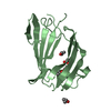
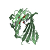
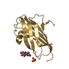
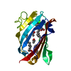
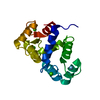
| Entry | Database: PDB / ID: 1pu5 | ||||||
|---|---|---|---|---|---|---|---|
| Title | GM2-activator Protein crystal structure | ||||||
 Components Components | Ganglioside GM2 activator | ||||||
 Keywords Keywords | LIPID BINDING PROTEIN / Beta cup / large lipid binding pocket / protein dynamics | ||||||
| Function / homology |  Function and homology information Function and homology informationsphingolipid activator protein activity / beta-N-acetylgalactosaminidase activity / glycosphingolipid catabolic process / lipid carrier activity / Glycosphingolipid catabolism / lipid storage / ganglioside catabolic process / oligosaccharide catabolic process / neuromuscular process controlling balance / phospholipase activator activity ...sphingolipid activator protein activity / beta-N-acetylgalactosaminidase activity / glycosphingolipid catabolic process / lipid carrier activity / Glycosphingolipid catabolism / lipid storage / ganglioside catabolic process / oligosaccharide catabolic process / neuromuscular process controlling balance / phospholipase activator activity / lipid transport / lysosomal lumen / cytoplasmic side of plasma membrane / azurophil granule lumen / basolateral plasma membrane / learning or memory / apical plasma membrane / Neutrophil degranulation / extracellular exosome / extracellular region / cytosol Similarity search - Function | ||||||
| Biological species |  Homo sapiens (human) Homo sapiens (human) | ||||||
| Method |  X-RAY DIFFRACTION / SYNCHROTRON / MOLECULAR REPLACEMENT / Resolution: 1.9 Å X-RAY DIFFRACTION / SYNCHROTRON / MOLECULAR REPLACEMENT / Resolution: 1.9 Å | ||||||
 Authors Authors | Wright, C.S. / Zhao, Q. / Rastinejad, F. | ||||||
 Citation Citation | Journal: J.Mol.Biol. / Year: 2003 Title: Structural analysis of lipid complexes of GM2-activator protein. Authors: Wright, C.S. / Zhao, Q. / Rastinejad, F. | ||||||
| History |
|
- Structure visualization
| Structure viewer | Molecule: MolmilJmol/JSmol |
|---|
- Downloads & links
-Download
| PDBx/mmCIF format | 1pu5.cif.gz | 113.3 KB | Display | PDBx/mmCIF format |
|---|---|---|---|---|
| PDB format | pdb1pu5.ent.gz | 88.4 KB | Display | PDB format |
| PDBx/mmJSON format | 1pu5.json.gz | Tree view | PDBx/mmJSON format | |
| Others |  Other downloads Other downloads |
-Validation report
| Arichive directory | https://data.pdbj.org/pub/pdb/validation_reports/pu/1pu5ftp://data.pdbj.org/pub/pdb/validation_reports/pu/1pu5 | HTTPS FTP |
|---|
-Related structure data
| Related structure data | 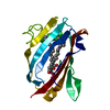 1pubC  1g13S S: Starting model for refinement C: citing same article ( |
|---|---|
| Similar structure data |

-Links
 PDBj
PDBj
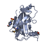
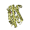
- Assembly
| Deposited unit | 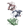
| ||||||||
|---|---|---|---|---|---|---|---|---|---|
| 1 | 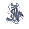
| ||||||||
| 2 | 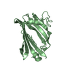
| ||||||||
| 3 | 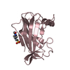
| ||||||||
| 4 | 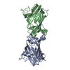
| ||||||||
| Unit cell |
|
-Components
| #1: Protein | Mass: 17827.557 Da / Num. of mol.: 3 Source method: isolated from a genetically manipulated source Source: (gene. exp.) Homo sapiens (human) / Gene: GM2A / Plasmid: pET16b (Novagen) / Species (production host): Escherichia coli / Production host:  #2: Chemical |   Mass: 238.305 Da / Num. of mol.: 2 / Source method: obtained synthetically / Formula: C8H18N2O4S / Comment: pH buffer*YM Mass: 238.305 Da / Num. of mol.: 2 / Source method: obtained synthetically / Formula: C8H18N2O4S / Comment: pH buffer*YM#3: Water | ChemComp-HOH / |  Mass: 18.015 Da / Num. of mol.: 432 / Source method: isolated from a natural source / Formula: H2O Mass: 18.015 Da / Num. of mol.: 432 / Source method: isolated from a natural source / Formula: H2OHas protein modification | Y | |
|---|
-Experimental details
-Experiment
| Experiment | Method: X-RAY DIFFRACTION / Number of used crystals: 1 |
|---|
- Sample preparation
| Crystal | Density Matthews: 3.06 Å3/Da / Density % sol: 59.83 % |
|---|---|
| Crystal grow | Temperature: 278 K / Method: vapor diffusion, hanging drop / pH: 7.7 Details: Peg 4000, iso-propanol, Hepes buffer, pH 7.7, VAPOR DIFFUSION, HANGING DROP, temperature 278.0K |
-Data collection
| Diffraction | Mean temperature: 123 K |
|---|---|
| Diffraction source | Source: SYNCHROTRON / Site: APS  / Beamline: 17-BM / Wavelength: 0.979 Å / Beamline: 17-BM / Wavelength: 0.979 Å |
| Radiation | Protocol: SINGLE WAVELENGTH / Monochromatic (M) / Laue (L): M / Scattering type: x-ray |
| Radiation wavelength | Wavelength: 0.979 Å / Relative weight: 1 |
| Reflection | Resolution: 1.9→50 Å / Num. all: 51764 / Num. obs: 50312 / % possible obs: 97.2 % / Observed criterion σ(F): 1 / Observed criterion σ(I): 1 / Redundancy: 3.5 % / Biso Wilson estimate: 22.8 Å2 / Rmerge(I) obs: 0.049 / Rsym value: 0.049 / Net I/σ(I): 5 |
| Reflection shell | Resolution: 1.9→1.97 Å / Rmerge(I) obs: 0.515 |
- Processing
| Software |
| |||||||||||||||||||||||||
|---|---|---|---|---|---|---|---|---|---|---|---|---|---|---|---|---|---|---|---|---|---|---|---|---|---|---|
| Refinement | Method to determine structure: MOLECULAR REPLACEMENT Starting model: PDB entry 1G13 Resolution: 1.9→7.99 Å / Rfactor Rfree error: 0.004 / Data cutoff high absF: 2018589.88 / Data cutoff high rms absF: 2018589.88 / Data cutoff low absF: 0 / Isotropic thermal model: RESTRAINED / Cross valid method: THROUGHOUT / σ(F): 1 / σ(I): 1 / Stereochemistry target values: Engh & Huber
| |||||||||||||||||||||||||
| Solvent computation | Solvent model: FLAT MODEL / Bsol: 92.897 Å2 / ksol: 0.512718 e/Å3 | |||||||||||||||||||||||||
| Displacement parameters | Biso mean: 30.5 Å2
| |||||||||||||||||||||||||
| Refine analyze |
| |||||||||||||||||||||||||
| Refinement step | Cycle: LAST / Resolution: 1.9→7.99 Å
| |||||||||||||||||||||||||
| Refine LS restraints |
| |||||||||||||||||||||||||
| LS refinement shell | Resolution: 1.9→2.02 Å / Rfactor Rfree error: 0.01 / Total num. of bins used: 6
| |||||||||||||||||||||||||
| Xplor file |
|
