Resolution: 1→1.01 Å / Redundancy: 4 % / Mean I/σ(I) obs: 3.8 / Rsym value: 0.385 / % possible all: 92.6
Reflection
*PLUS
Highest resolution: 1 Å / Lowest resolution: 22 Å / Num. obs: 86663 / Rmerge(I) obs: 0.073
-
Processing
Software
Name
Classification
SHELXL
refinement
DENZO
datareduction
SCALEPACK
datascaling
Refinement
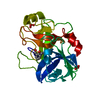
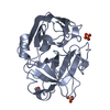
Method to determine structure: MOLECULAR REPLACEMENT / Resolution: 1→20 Å / Num. parameters: 19056 / Num. restraintsaints: 25644 / Stereochemistry target values: Engh & Huber Details: ANISOTROPIC REFINEMENT. Initially Rfree (building stage) was used (10% of data at random), later none. There is a peptide chain C, GLY-GLY-ARG. The ARG is in two alternate conformations, A ...Details: ANISOTROPIC REFINEMENT. Initially Rfree (building stage) was used (10% of data at random), later none. There is a peptide chain C, GLY-GLY-ARG. The ARG is in two alternate conformations, A and B. There is a LYS 403 ligand that also occupies the same space as the ARG and is not part of the peptide chain. The LYS is labelled as alternate conformation C.
Rfactor
Num. reflection
% reflection
Rwork
0.1279
-
-
all
-
86657
-
obs
-
86657
94.9 %
Refine analyze
Num. disordered residues: 55 / Occupancy sum hydrogen: 1499.14 / Occupancy sum non hydrogen: 1883.6
In the structure databanks used in Yorodumi, some data are registered as the other names, "COVID-19 virus" and "2019-nCoV". Here are the details of the virus and the list of structure data.
Jan 31, 2019. EMDB accession codes are about to change! (news from PDBe EMDB page)
EMDB accession codes are about to change! (news from PDBe EMDB page)
The allocation of 4 digits for EMDB accession codes will soon come to an end. Whilst these codes will remain in use, new EMDB accession codes will include an additional digit and will expand incrementally as the available range of codes is exhausted. The current 4-digit format prefixed with “EMD-” (i.e. EMD-XXXX) will advance to a 5-digit format (i.e. EMD-XXXXX), and so on. It is currently estimated that the 4-digit codes will be depleted around Spring 2019, at which point the 5-digit format will come into force.
The EM Navigator/Yorodumi systems omit the EMD- prefix.
Related info.:Q: What is EMD? / ID/Accession-code notation in Yorodumi/EM Navigator
Yorodumi is a browser for structure data from EMDB, PDB, SASBDB, etc.
This page is also the successor to EM Navigator detail page, and also detail information page/front-end page for Omokage search.
The word "yorodu" (or yorozu) is an old Japanese word meaning "ten thousand". "mi" (miru) is to see.
Related info.:EMDB / PDB / SASBDB / Comparison of 3 databanks / Yorodumi Search / Aug 31, 2016. New EM Navigator & Yorodumi / Yorodumi Papers / Jmol/JSmol / Function and homology information / Changes in new EM Navigator and Yorodumi
 Movie
Movie Controller


 Open data
 Basic information
 Components
 Keywords
 Function and homology information
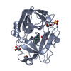
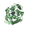
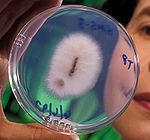 Fusarium oxysporum (fungus)
 X-RAY DIFFRACTION /
  Authors
 Citation
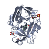
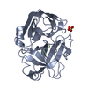
 Structure visualization
 Downloads & links
 Other downloads
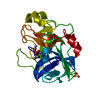


 PDBj
PDBj

 Assembly
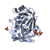


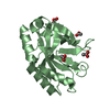
 Mass: 96.063 Da / Num. of mol.: 3 / Source method: obtained synthetically / Formula: SO4
 Type: L-peptide linking / Mass: 147.195 Da / Num. of mol.: 1 / Source method: obtained synthetically / Formula: C6H15N2O2
 Mass: 192.124 Da / Num. of mol.: 1 / Source method: obtained synthetically / Formula: C6H8O7
 Sample preparation
 / Beamline: X13 / Wavelength: 0.802 Å
 Processing