 Movie
Movie Controller

[English] 日本語
 Yorodumi
- PDB-1pnd: ACCURACY AND PRECISION IN PROTEIN CRYSTAL STRUCTURE ANALYSIS: TWO... -
+ Open data

- Basic information
| Entry | Database: PDB / ID: 1pnd | ||||||
|---|---|---|---|---|---|---|---|
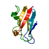
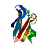
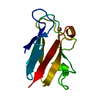
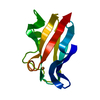
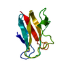
| Title | ACCURACY AND PRECISION IN PROTEIN CRYSTAL STRUCTURE ANALYSIS: TWO INDEPENDENT REFINEMENTS OF THE STRUCTURE OF POPLAR PLASTOCYANIN AT 173K | ||||||
 Components Components | PLASTOCYANIN | ||||||
 Keywords Keywords | ELECTRON TRANSPORT | ||||||
| Function / homology |  Function and homology information Function and homology informationchloroplast thylakoid lumen / chloroplast thylakoid membrane / electron transfer activity / copper ion binding Similarity search - Function | ||||||
| Biological species |  | ||||||
| Method |  X-RAY DIFFRACTION / SYNCHROTRON / Resolution: 1.6 Å X-RAY DIFFRACTION / SYNCHROTRON / Resolution: 1.6 Å | ||||||
 Authors Authors | Fields, B.A. / Guss, J.M. / Freeman, H.C. | ||||||
 Citation Citation | Journal: Acta Crystallogr.,Sect.D / Year: 1994 Title: Accuracy and precision in protein crystal structure analysis: two independent refinements of the structure of poplar plastocyanin at 173 K. Authors: Fields, B.A. / Bartsch, H.H. / Bartunik, H.D. / Cordes, F. / Guss, J.M. / Freeman, H.C. #1: Journal: Acta Crystallogr.,Sect.B / Year: 1992Title: Accuracy and Precision in Protein Structure Analysis: Restrained Least-Squares Refinement of the Structure of Poplar Plastocyanin at 1.33 Angstroms Resolution Authors: Guss, J.M. / Bartunik, H.D. / Freeman, H.C. #2: Journal: J.Mol.Biol. / Year: 1983Title: Structure of Oxidised Poplar Plastocyanin at 1.6 Angstroms Resolution Authors: Guss, J.M. / Freeman, H.C. #3: Journal: Nature / Year: 1978Title: X-Ray Crystal Structure Analysis of Plastocyanin at 2.7 Angstroms Resolution Authors: Colman, P.M. / Freeman, H.C. / Guss, J.M. / Murata, M. / Norris, V.A. / Ramshaw, J.A.M. / Venkatappa, M.P. / Freeman, H.C. #4: Journal: J.Mol.Biol. / Year: 1977Title: Preliminary Crystallographic Data for a Copper Containing Protein, Plastocyanin Authors: Chapman, G.V. / Colman, P.M. / Freeman, H.C. / Guss, J.M. / Murata, M. / Norris, V.A. / Ramashaw, J.A.M. / Venkatappa, M.P. / Freeman, H.C. | ||||||
| History |
|
- Structure visualization
| Structure viewer | Molecule: MolmilJmol/JSmol |
|---|
- Downloads & links
-Download
| PDBx/mmCIF format | 1pnd.cif.gz | 34.3 KB | Display | PDBx/mmCIF format |
|---|---|---|---|---|
| PDB format | pdb1pnd.ent.gz | 22.5 KB | Display | PDB format |
| PDBx/mmJSON format | 1pnd.json.gz | Tree view | PDBx/mmJSON format | |
| Others |  Other downloads Other downloads |
-Validation report
| Arichive directory | https://data.pdbj.org/pub/pdb/validation_reports/pn/1pndftp://data.pdbj.org/pub/pdb/validation_reports/pn/1pnd | HTTPS FTP |
|---|
-Related structure data


-Links
 PDBj
PDBj- Assembly
| Deposited unit | 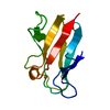
| ||||||||
|---|---|---|---|---|---|---|---|---|---|
| 1 |
| ||||||||
| Unit cell |
| ||||||||
| Atom site foot note | 1: CIS PROLINE - PRO 16 / 2: CIS PROLINE - PRO 36 3: THE LIGANDS OF THE CU ATOM ARE ND1 HIS 37, SG CYS 84, ND1 HIS 87, AND SD MET 92. |
-Components
| #1: Protein | Mass: 10493.607 Da / Num. of mol.: 1 Source method: isolated from a genetically manipulated source Source: (gene. exp.) |
|---|---|
| #2: Chemical | ChemComp-CU /   Mass: 63.546 Da / Num. of mol.: 1 / Source method: obtained synthetically / Formula: Cu Mass: 63.546 Da / Num. of mol.: 1 / Source method: obtained synthetically / Formula: Cu |
| #3: Water | ChemComp-HOH /  Mass: 18.015 Da / Num. of mol.: 171 / Source method: isolated from a natural source / Formula: H2O Mass: 18.015 Da / Num. of mol.: 171 / Source method: isolated from a natural source / Formula: H2O |
-Experimental details
-Experiment
| Experiment | Method: X-RAY DIFFRACTION |
|---|
- Sample preparation
| Crystal | Density Matthews: 1.82 Å3/Da / Density % sol: 32.48 % | ||||||||||||||||||||
|---|---|---|---|---|---|---|---|---|---|---|---|---|---|---|---|---|---|---|---|---|---|
| Crystal grow | *PLUS pH: 6 / Method: vapor diffusionDetails: taken from Chapman, G.V. et al (1977). J. Mol. Biol., 110, 187-189. | ||||||||||||||||||||
| Components of the solutions | *PLUS
|
-Data collection
| Diffraction source | Source: SYNCHROTRON / Site: EMBL/DESY, HAMBURG  / Beamline: X31 / Beamline: X31 |
|---|---|
| Detector | Type: ENRAF-NONIUS / Detector: AREA DETECTOR |
| Radiation | Scattering type: x-ray |
| Radiation wavelength | Relative weight: 1 |
- Processing
| Software |
| ||||||||||||||||||||||||||||||||||||||||||||||||||||||||||||
|---|---|---|---|---|---|---|---|---|---|---|---|---|---|---|---|---|---|---|---|---|---|---|---|---|---|---|---|---|---|---|---|---|---|---|---|---|---|---|---|---|---|---|---|---|---|---|---|---|---|---|---|---|---|---|---|---|---|---|---|---|---|
| Refinement | Resolution: 1.6→6 Å Details: THE STRUCTURE WAS REFINED INDEPENDENTLY IN SYDNEY USING PROLSQ AND IN HAMBURG USING EREF. THE STRUCTURES WERE COMPARED ONLY AFTER THE REFINEMENTS WERE COMPLETE.
| ||||||||||||||||||||||||||||||||||||||||||||||||||||||||||||
| Refinement step | Cycle: LAST / Resolution: 1.6→6 Å
| ||||||||||||||||||||||||||||||||||||||||||||||||||||||||||||
| Refine LS restraints |
| ||||||||||||||||||||||||||||||||||||||||||||||||||||||||||||
| Software | *PLUS Name: EREF / Classification: refinement | ||||||||||||||||||||||||||||||||||||||||||||||||||||||||||||
| Refinement | *PLUS Highest resolution: 1.6 Å / Lowest resolution: 6 Å / Num. reflection obs: 7393 / Rfactor obs: 0.135 | ||||||||||||||||||||||||||||||||||||||||||||||||||||||||||||
| Solvent computation | *PLUS | ||||||||||||||||||||||||||||||||||||||||||||||||||||||||||||
| Displacement parameters | *PLUS Biso mean: 11 Å2 |
