 Movie
Movie Controller

[English] 日本語
 Yorodumi
- PDB-1pn4: Crystal structure of 2-enoyl-CoA hydratase 2 domain of Candida tr... -
+ Open data

- Basic information
| Entry | Database: PDB / ID: 1pn4 | ||||||
|---|---|---|---|---|---|---|---|
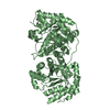
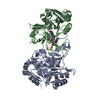
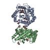
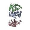
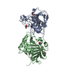
| Title | Crystal structure of 2-enoyl-CoA hydratase 2 domain of Candida tropicalis multifunctional enzyme type 2 complexed with (3R)-hydroxydecanoyl-CoA. | ||||||
 Components Components | Peroxisomal hydratase-dehydrogenase-epimerase | ||||||
 Keywords Keywords | LYASE / Hot-Dog fold / Hydratase 2 motif / Oxyanion hole / Enzyme-product complex | ||||||
| Function / homology |  Function and homology information Function and homology information: / (3R)-3-hydroxyacyl-CoA dehydrogenase (NAD+) activity / enoyl-CoA hydratase 2 / 3-hydroxyacyl-CoA dehydratase activity / (3S)-3-hydroxyacyl-CoA dehydrogenase (NAD+) activity / fatty acid beta-oxidation / isomerase activity / peroxisome Similarity search - Function | ||||||
| Biological species |  Candida tropicalis (yeast) Candida tropicalis (yeast) | ||||||
| Method |  X-RAY DIFFRACTION / SYNCHROTRON / MOLECULAR REPLACEMENT / Resolution: 2.35 Å X-RAY DIFFRACTION / SYNCHROTRON / MOLECULAR REPLACEMENT / Resolution: 2.35 Å | ||||||
 Authors Authors | Koski, M.K. / Haapalainen, A.M. / Hiltunen, J.K. / Glumoff, T. | ||||||
 Citation Citation | Journal: J.Biol.Chem. / Year: 2004 Title: A Two-domain Structure of One Subunit Explains Unique Features of Eukaryotic Hydratase 2. Authors: Koski, M.K. / Haapalainen, A.M. / Hiltunen, J.K. / Glumoff, T. | ||||||
| History |
|
- Structure visualization
| Structure viewer | Molecule: MolmilJmol/JSmol |
|---|
- Downloads & links
-Download
| PDBx/mmCIF format | 1pn4.cif.gz | 232.6 KB | Display | PDBx/mmCIF format |
|---|---|---|---|---|
| PDB format | pdb1pn4.ent.gz | 184.8 KB | Display | PDB format |
| PDBx/mmJSON format | 1pn4.json.gz | Tree view | PDBx/mmJSON format | |
| Others |  Other downloads Other downloads |
-Validation report
| Arichive directory | https://data.pdbj.org/pub/pdb/validation_reports/pn/1pn4ftp://data.pdbj.org/pub/pdb/validation_reports/pn/1pn4 | HTTPS FTP |
|---|
-Related structure data
| Related structure data |  1pn2SC S: Starting model for refinement C: citing same article ( |
|---|---|
| Similar structure data |


-Links
 PDBj
PDBj


- Assembly
| Deposited unit | 
| ||||||||
|---|---|---|---|---|---|---|---|---|---|
| 1 | 
| ||||||||
| 2 | 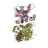
| ||||||||
| Unit cell |
| ||||||||
| Details | The asymmetric unit is complete and the biological unit is a dimer. |
-Components
| #1: Protein | Mass: 31396.729 Da / Num. of mol.: 4 / Fragment: 2-enoyl-CoA hydratase 2 domain / Mutation: E627M, H813Q Source method: isolated from a genetically manipulated source Source: (gene. exp.) Candida tropicalis (yeast) / Gene: FOX2 / Plasmid: pET3A / Species (production host): Escherichia coli / Production host:  References: UniProt: P22414, Lyases; Carbon-oxygen lyases; Hydro-lyases #2: Chemical |   Mass: 937.783 Da / Num. of mol.: 3 / Source method: obtained synthetically / Formula: C31H54N7O18P3S Mass: 937.783 Da / Num. of mol.: 3 / Source method: obtained synthetically / Formula: C31H54N7O18P3S#3: Chemical | ChemComp-EDO /   Mass: 62.068 Da / Num. of mol.: 4 / Source method: obtained synthetically / Formula: C2H6O2 Mass: 62.068 Da / Num. of mol.: 4 / Source method: obtained synthetically / Formula: C2H6O2#4: Water | ChemComp-HOH / |  Mass: 18.015 Da / Num. of mol.: 465 / Source method: isolated from a natural source / Formula: H2O Mass: 18.015 Da / Num. of mol.: 465 / Source method: isolated from a natural source / Formula: H2O |
|---|
-Experimental details
-Experiment
| Experiment | Method: X-RAY DIFFRACTION / Number of used crystals: 1 |
|---|
- Sample preparation
| Crystal | Density Matthews: 2.39 Å3/Da / Density % sol: 48.53 % |
|---|---|
| Crystal grow | Temperature: 277 K / Method: vapor diffusion, hanging drop / pH: 7 Details: PEG 2000 MME, HEPES, trans-2-decenoyl-CoA, pH 7.0, VAPOR DIFFUSION, HANGING DROP, temperature 277K |
-Data collection
| Diffraction | Mean temperature: 100 K |
|---|---|
| Diffraction source | Source: SYNCHROTRON / Site: EMBL/DESY, HAMBURG  / Beamline: X13 / Wavelength: 0.8019 Å / Beamline: X13 / Wavelength: 0.8019 Å |
| Detector | Type: MAR CCD 165 mm / Detector: CCD / Date: Aug 9, 2002 |
| Radiation | Monochromator: TRIANGULAR MONOCHROMATOR / Protocol: SINGLE WAVELENGTH / Monochromatic (M) / Laue (L): M / Scattering type: x-ray |
| Radiation wavelength | Wavelength: 0.8019 Å / Relative weight: 1 |
| Reflection | Resolution: 2.35→35 Å / Num. all: 49040 / Num. obs: 47255 / % possible obs: 96.4 % / Observed criterion σ(F): 0 / Observed criterion σ(I): 0 / Redundancy: 3 % / Biso Wilson estimate: 30.14 Å2 / Rmerge(I) obs: 0.064 / Net I/σ(I): 12.1 |
| Reflection shell | Resolution: 2.35→2.5 Å / Redundancy: 2.9 % / Rmerge(I) obs: 0.232 / Mean I/σ(I) obs: 4.53 / Num. unique all: 7461 / % possible all: 90.1 |
- Processing
| Software |
| |||||||||||||||||||||||||
|---|---|---|---|---|---|---|---|---|---|---|---|---|---|---|---|---|---|---|---|---|---|---|---|---|---|---|
| Refinement | Method to determine structure: MOLECULAR REPLACEMENT Starting model: Subunit D of PDB ENTRY 1PN2 Resolution: 2.35→35 Å / Cross valid method: THROUGHOUT / σ(F): 0 / σ(I): 0 / Stereochemistry target values: Engh & Huber
| |||||||||||||||||||||||||
| Refinement step | Cycle: LAST / Resolution: 2.35→35 Å
| |||||||||||||||||||||||||
| Refine LS restraints |
|
