 Movie
Movie Controller

[English] 日本語
 Yorodumi
- PDB-1pjh: Structural studies on delta3-delta2-enoyl-CoA isomerase: the vari... -
+ Open data

- Basic information
| Entry | Database: PDB / ID: 1pjh | ||||||
|---|---|---|---|---|---|---|---|
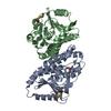
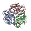
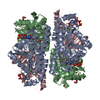
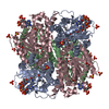
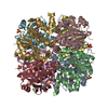
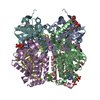
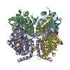
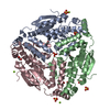
| Title | Structural studies on delta3-delta2-enoyl-CoA isomerase: the variable mode of assembly of the trimeric disks of the crotonase superfamily | ||||||
 Components Components | enoyl-CoA isomerase; Eci1p | ||||||
 Keywords Keywords | ISOMERASE / beta-beta-alpha spiral fold / inter-trimer contacts | ||||||
| Function / homology |  Function and homology information Function and homology informationBeta-oxidation of very long chain fatty acids / Delta3-Delta2-enoyl-CoA isomerase / delta(3)-delta(2)-enoyl-CoA isomerase activity / Peroxisomal protein import / fatty acid beta-oxidation / peroxisomal matrix / peroxisome Similarity search - Function | ||||||
| Biological species |  | ||||||
| Method |  X-RAY DIFFRACTION / SYNCHROTRON / MOLECULAR REPLACEMENT / Resolution: 2.1 Å X-RAY DIFFRACTION / SYNCHROTRON / MOLECULAR REPLACEMENT / Resolution: 2.1 Å | ||||||
 Authors Authors | Mursula, A.M. / Hiltunen, J.K. / Wierenga, R.K. | ||||||
 Citation Citation | Journal: Febs Lett. / Year: 2004 Title: Structural studies on delta(3)-delta(2)-enoyl-CoA isomerase: the variable mode of assembly of the trimeric disks of the crotonase superfamily. Authors: Mursula, A.M. / Hiltunen, J.K. / Wierenga, R.K. | ||||||
| History |
|
- Structure visualization
| Structure viewer | Molecule: MolmilJmol/JSmol |
|---|
- Downloads & links
-Download
| PDBx/mmCIF format | 1pjh.cif.gz | 171.4 KB | Display | PDBx/mmCIF format |
|---|---|---|---|---|
| PDB format | pdb1pjh.ent.gz | 136.5 KB | Display | PDB format |
| PDBx/mmJSON format | 1pjh.json.gz | Tree view | PDBx/mmJSON format | |
| Others |  Other downloads Other downloads |
-Validation report
| Arichive directory | https://data.pdbj.org/pub/pdb/validation_reports/pj/1pjhftp://data.pdbj.org/pub/pdb/validation_reports/pj/1pjh | HTTPS FTP |
|---|
-Related structure data
| Related structure data |  1hnuS S: Starting model for refinement |
|---|---|
| Similar structure data |

-Links
 PDBj
PDBj
- Assembly
| Deposited unit | 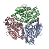
| ||||||||
|---|---|---|---|---|---|---|---|---|---|
| 1 | 
| ||||||||
| Unit cell |
|
-Components
| #1: Protein | Mass: 31718.369 Da / Num. of mol.: 3 Source method: isolated from a genetically manipulated source Source: (gene. exp.) Gene: ECI1 / Plasmid: pET3a / Production host:  References: UniProt: Q05871, Delta3-Delta2-enoyl-CoA isomerase #2: Chemical | ChemComp-SO4 /   Mass: 96.063 Da / Num. of mol.: 10 / Source method: obtained synthetically / Formula: SO4 Mass: 96.063 Da / Num. of mol.: 10 / Source method: obtained synthetically / Formula: SO4#3: Chemical | ChemComp-GOL /   Mass: 92.094 Da / Num. of mol.: 4 / Source method: obtained synthetically / Formula: C3H8O3 Mass: 92.094 Da / Num. of mol.: 4 / Source method: obtained synthetically / Formula: C3H8O3#4: Water | ChemComp-HOH / |  Mass: 18.015 Da / Num. of mol.: 382 / Source method: isolated from a natural source / Formula: H2O Mass: 18.015 Da / Num. of mol.: 382 / Source method: isolated from a natural source / Formula: H2O |
|---|
-Experimental details
-Experiment
| Experiment | Method: X-RAY DIFFRACTION / Number of used crystals: 1 |
|---|
- Sample preparation
| Crystal | Density Matthews: 3.84 Å3/Da / Density % sol: 67.95 % | ||||||||||||||||||||||||||||||||||||||||||||||||||||||||||||||||||||||
|---|---|---|---|---|---|---|---|---|---|---|---|---|---|---|---|---|---|---|---|---|---|---|---|---|---|---|---|---|---|---|---|---|---|---|---|---|---|---|---|---|---|---|---|---|---|---|---|---|---|---|---|---|---|---|---|---|---|---|---|---|---|---|---|---|---|---|---|---|---|---|---|
| Crystal grow | Temperature: 295 K / Method: vapor diffusion, hanging drop / pH: 7 Details: ADA/NaOH, magnesium sulphate, ammonium sulphate, pH 7.0, VAPOR DIFFUSION, HANGING DROP, temperature 295K | ||||||||||||||||||||||||||||||||||||||||||||||||||||||||||||||||||||||
| Crystal grow | *PLUS Temperature: 295 K / pH: 7.2 / Method: vapor diffusion, hanging drop | ||||||||||||||||||||||||||||||||||||||||||||||||||||||||||||||||||||||
| Components of the solutions | *PLUS
|
-Data collection
| Diffraction | Mean temperature: 100 K |
|---|---|
| Diffraction source | Source: SYNCHROTRON / Site: EMBL/DESY, HAMBURG  / Beamline: X11 / Wavelength: 0.801 Å / Beamline: X11 / Wavelength: 0.801 Å |
| Detector | Type: MARRESEARCH / Detector: CCD / Date: Nov 10, 2002 |
| Radiation | Monochromator: triangular monochromator and bent mirror / Protocol: SINGLE WAVELENGTH / Monochromatic (M) / Laue (L): M / Scattering type: x-ray |
| Radiation wavelength | Wavelength: 0.801 Å / Relative weight: 1 |
| Reflection | Resolution: 2.1→20 Å / Num. all: 86026 / Num. obs: 86026 / % possible obs: 98.9 % / Observed criterion σ(F): 2 / Observed criterion σ(I): 2 / Redundancy: 8 % / Biso Wilson estimate: 37.4 Å2 / Rmerge(I) obs: 0.04 / Net I/σ(I): 28.5 |
| Reflection shell | Resolution: 2.1→2.2 Å / Rmerge(I) obs: 0.199 / Mean I/σ(I) obs: 10.5 / Num. unique all: 10418 / % possible all: 93.4 |
| Reflection | *PLUS Redundancy: 8 % / Num. measured all: 690091 / Rmerge(I) obs: 0.04 |
| Reflection shell | *PLUS % possible obs: 93.4 % |
- Processing
| Software |
| ||||||||||||||||||||
|---|---|---|---|---|---|---|---|---|---|---|---|---|---|---|---|---|---|---|---|---|---|
| Refinement | Method to determine structure: MOLECULAR REPLACEMENT Starting model: 1HNU Resolution: 2.1→20 Å / σ(F): 2 / Stereochemistry target values: Engh & Huber
| ||||||||||||||||||||
| Refinement step | Cycle: LAST / Resolution: 2.1→20 Å
| ||||||||||||||||||||
| Refine LS restraints |
| ||||||||||||||||||||
| Software | *PLUS Version: 5 / Classification: refinement | ||||||||||||||||||||
| Refinement | *PLUS Num. reflection obs: 81748 / Num. reflection Rfree: 4278 | ||||||||||||||||||||
| Solvent computation | *PLUS | ||||||||||||||||||||
| Displacement parameters | *PLUS | ||||||||||||||||||||
| Refine LS restraints | *PLUS
|
