 Movie
Movie Controller

[English] 日本語
 Yorodumi
- PDB-1pix: Crystal structure of the carboxyltransferase subunit of the bacte... -
+ Open data

- Basic information
| Entry | Database: PDB / ID: 1pix | ||||||
|---|---|---|---|---|---|---|---|
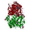
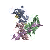
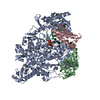
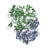
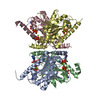
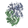
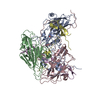
| Title | Crystal structure of the carboxyltransferase subunit of the bacterial ion pump glutaconyl-coenzyme A decarboxylase | ||||||
 Components Components | Glutaconyl-CoA decarboxylase A subunit | ||||||
 Keywords Keywords | LYASE / decarboxylase / biotin-dependent ion pump / carboxyltransferase | ||||||
| Function / homology |  Function and homology information Function and homology informationglutaconyl-CoA decarboxylase / glutaconyl-CoA decarboxylase activity / : / methylcrotonoyl-CoA carboxylase complex / methylcrotonoyl-CoA carboxylase activity / L-leucine catabolic process / sodium ion transport Similarity search - Function | ||||||
| Biological species |  Acidaminococcus fermentans (bacteria) Acidaminococcus fermentans (bacteria) | ||||||
| Method |  X-RAY DIFFRACTION / SYNCHROTRON / MAD / Resolution: 2.2 Å X-RAY DIFFRACTION / SYNCHROTRON / MAD / Resolution: 2.2 Å | ||||||
 Authors Authors | Wendt, K.S. / Schall, I. / Huber, R. / Buckel, W. / Jacob, U. | ||||||
 Citation Citation | Journal: Embo J. / Year: 2003 Title: Crystal structure of the carboxyltransferase subunit of the bacterial sodium ion pump glutaconyl-coenzyme A decarboxylase Authors: Wendt, K.S. / Schall, I. / Huber, R. / Buckel, W. / Jacob, U. | ||||||
| History |
|
- Structure visualization
| Structure viewer | Molecule: MolmilJmol/JSmol |
|---|
- Downloads & links
-Download
| PDBx/mmCIF format | 1pix.cif.gz | 245.1 KB | Display | PDBx/mmCIF format |
|---|---|---|---|---|
| PDB format | pdb1pix.ent.gz | 197.8 KB | Display | PDB format |
| PDBx/mmJSON format | 1pix.json.gz | Tree view | PDBx/mmJSON format | |
| Others |  Other downloads Other downloads |
-Validation report
| Arichive directory | https://data.pdbj.org/pub/pdb/validation_reports/pi/1pixftp://data.pdbj.org/pub/pdb/validation_reports/pi/1pix | HTTPS FTP |
|---|
-Related structure data
| Similar structure data |
|---|

-Links
 PDBj
PDBj- Assembly
| Deposited unit | 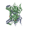
| ||||||||
|---|---|---|---|---|---|---|---|---|---|
| 1 |
| ||||||||
| 2 | 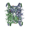
| ||||||||
| Unit cell |
| ||||||||
| Details | The biological assembly is generated by the two monomers in the asymmetric unit |
-Components
| #1: Protein | Mass: 64423.508 Da / Num. of mol.: 2 Source method: isolated from a genetically manipulated source Source: (gene. exp.) Acidaminococcus fermentans (bacteria) / Gene: GCDA / Plasmid: pJF118HE / Production host: #2: Chemical | ChemComp-SO4 /   Mass: 96.063 Da / Num. of mol.: 5 / Source method: obtained synthetically / Formula: SO4 Mass: 96.063 Da / Num. of mol.: 5 / Source method: obtained synthetically / Formula: SO4#3: Chemical | ChemComp-FMT /   Mass: 46.025 Da / Num. of mol.: 4 / Source method: obtained synthetically / Formula: CH2O2 Mass: 46.025 Da / Num. of mol.: 4 / Source method: obtained synthetically / Formula: CH2O2#4: Water | ChemComp-HOH / |  Mass: 18.015 Da / Num. of mol.: 661 / Source method: isolated from a natural source / Formula: H2O Mass: 18.015 Da / Num. of mol.: 661 / Source method: isolated from a natural source / Formula: H2O |
|---|
-Experimental details
-Experiment
| Experiment | Method: X-RAY DIFFRACTION / Number of used crystals: 1 |
|---|
- Sample preparation
| Crystal | Density Matthews: 3.65 Å3/Da / Density % sol: 66.26 % | |||||||||||||||||||||||||||||||||||
|---|---|---|---|---|---|---|---|---|---|---|---|---|---|---|---|---|---|---|---|---|---|---|---|---|---|---|---|---|---|---|---|---|---|---|---|---|
| Crystal grow | Temperature: 298 K / Method: vapor diffusion, sitting drop / pH: 4.7 Details: acetate buffer, sodium formate, pH 4.7, VAPOR DIFFUSION, SITTING DROP, temperature 298K | |||||||||||||||||||||||||||||||||||
| Crystal grow | *PLUS pH: 7.5 / Method: vapor diffusion | |||||||||||||||||||||||||||||||||||
| Components of the solutions | *PLUS
|
-Data collection
| Diffraction | Mean temperature: 100 K |
|---|---|
| Diffraction source | Source: SYNCHROTRON / Site: MPG/DESY, HAMBURG  / Beamline: BW6 / Wavelength: 1.05 Å / Beamline: BW6 / Wavelength: 1.05 Å |
| Detector | Type: MARRESEARCH / Detector: CCD |
| Radiation | Protocol: SINGLE WAVELENGTH / Monochromatic (M) / Laue (L): M / Scattering type: x-ray |
| Radiation wavelength | Wavelength: 1.05 Å / Relative weight: 1 |
| Reflection | Resolution: 2.2→20 Å / Num. all: 96752 / Num. obs: 95460 / % possible obs: 98.7 % / Observed criterion σ(F): 2 / Observed criterion σ(I): 2 / Rmerge(I) obs: 0.069 |
| Reflection shell | Resolution: 2.2→2.21 Å / Rmerge(I) obs: 0.069 / Mean I/σ(I) obs: 13.7 / % possible all: 98.7 |
| Reflection | *PLUS Highest resolution: 2.2 Å / % possible obs: 99.1 % |
- Processing
| Software |
| |||||||||||||||||||||||||
|---|---|---|---|---|---|---|---|---|---|---|---|---|---|---|---|---|---|---|---|---|---|---|---|---|---|---|
| Refinement | Method to determine structure: MAD / Resolution: 2.2→20 Å / σ(F): 0 / Stereochemistry target values: Engh & Huber
| |||||||||||||||||||||||||
| Refinement step | Cycle: LAST / Resolution: 2.2→20 Å
| |||||||||||||||||||||||||
| Refinement | *PLUS Highest resolution: 2.2 Å / % reflection Rfree: 5 % | |||||||||||||||||||||||||
| Solvent computation | *PLUS | |||||||||||||||||||||||||
| Displacement parameters | *PLUS | |||||||||||||||||||||||||
| Refine LS restraints | *PLUS
|
