 Movie
Movie Controller

[English] 日本語
 Yorodumi
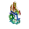
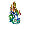
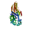
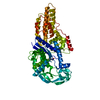
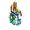
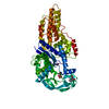
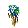
- PDB-1pfu: METHIONYL-TRNA SYNTHETASE FROM ESCHERICHIA COLI COMPLEXED WITH ME... -
+ Open data

- Basic information
| Entry | Database: PDB / ID: 1pfu | ||||||
|---|---|---|---|---|---|---|---|
| Title | METHIONYL-TRNA SYNTHETASE FROM ESCHERICHIA COLI COMPLEXED WITH METHIONINE PHOSPHINATE | ||||||
 Components Components | Methionyl-tRNA synthetase | ||||||
 Keywords Keywords | LIGASE / Rossmann fold | ||||||
| Function / homology |  Function and homology information Function and homology informationmethionine-tRNA ligase / methionine-tRNA ligase activity / methionyl-tRNA aminoacylation / cellular response to antibiotic / tRNA binding / protein homodimerization activity / zinc ion binding / ATP binding / membrane / cytosol Similarity search - Function | ||||||
| Biological species |  | ||||||
| Method |  X-RAY DIFFRACTION / SYNCHROTRON / MOLECULAR REPLACEMENT / Resolution: 1.91 Å X-RAY DIFFRACTION / SYNCHROTRON / MOLECULAR REPLACEMENT / Resolution: 1.91 Å | ||||||
 Authors Authors | Crepin, T. / Schmitt, E. / Mechulam, Y. / Sampson, P.B. / Vaughan, M.D. / Honek, J.F. / Blanquet, S. | ||||||
 Citation Citation | Journal: J.Mol.Biol. / Year: 2003 Title: Use of analogues of methionine and methionyl adenylate to sample conformational changes during catalysis in Escherichia coli methionyl-tRNA synthetase. Authors: Crepin, T. / Schmitt, E. / Mechulam, Y. / Sampson, P.B. / Vaughan, M.D. / Honek, J.F. / Blanquet, S. | ||||||
| History |
|
- Structure visualization
| Structure viewer | Molecule: MolmilJmol/JSmol |
|---|
- Downloads & links
-Download
| PDBx/mmCIF format | 1pfu.cif.gz | 131.1 KB | Display | PDBx/mmCIF format |
|---|---|---|---|---|
| PDB format | pdb1pfu.ent.gz | 100 KB | Display | PDB format |
| PDBx/mmJSON format | 1pfu.json.gz | Tree view | PDBx/mmJSON format | |
| Others |  Other downloads Other downloads |
-Validation report
| Arichive directory | https://data.pdbj.org/pub/pdb/validation_reports/pf/1pfuftp://data.pdbj.org/pub/pdb/validation_reports/pf/1pfu | HTTPS FTP |
|---|
-Related structure data
| Related structure data |  1p7pC  1pfvC  1pfwC  1pfyC  1pg0C  1pg2C  1qqtS C: citing same article ( S: Starting model for refinement |
|---|---|
| Similar structure data |


-Links
 PDBj
PDBj
- Assembly
| Deposited unit | 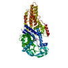
| ||||||||
|---|---|---|---|---|---|---|---|---|---|
| 1 |
| ||||||||
| Unit cell |
|
-Components
| #1: Protein | Mass: 62958.102 Da / Num. of mol.: 1 / Fragment: RESIDUES 1-551 Source method: isolated from a genetically manipulated source Source: (gene. exp.) |
|---|---|
| #2: Chemical | ChemComp-ZN /   Mass: 65.409 Da / Num. of mol.: 1 / Source method: obtained synthetically / Formula: Zn Mass: 65.409 Da / Num. of mol.: 1 / Source method: obtained synthetically / Formula: Zn |
| #3: Chemical | ChemComp-MPJ / (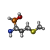  Type: L-peptide linking / Mass: 169.182 Da / Num. of mol.: 1 / Source method: obtained synthetically / Formula: C4H12NO2PS Type: L-peptide linking / Mass: 169.182 Da / Num. of mol.: 1 / Source method: obtained synthetically / Formula: C4H12NO2PS |
| #4: Water | ChemComp-HOH /  Mass: 18.015 Da / Num. of mol.: 443 / Source method: isolated from a natural source / Formula: H2O Mass: 18.015 Da / Num. of mol.: 443 / Source method: isolated from a natural source / Formula: H2O |
-Experimental details
-Experiment
| Experiment | Method: X-RAY DIFFRACTION / Number of used crystals: 1 |
|---|
- Sample preparation
| Crystal | Density Matthews: 2.35 Å3/Da / Density % sol: 47.69 % |
|---|---|
| Crystal grow | Temperature: 280 K / Method: vapor diffusion / pH: 7 Details: AMMONIUM CITRATE, POTASSIUM PHOSPHATE, pH 7, VAPOR DIFFUSION, temperature 280K |
| Crystal grow | *PLUS Method: unknown / Details: Mechulam, Y., (1999) J. Mol. Biol., 294, 1287. |
-Data collection
| Diffraction | Mean temperature: 100 K |
|---|---|
| Diffraction source | Source: SYNCHROTRON / Site: LURE  / Beamline: DW32 / Wavelength: 0.9674 Å / Beamline: DW32 / Wavelength: 0.9674 Å |
| Detector | Type: MARRESEARCH / Detector: IMAGE PLATE / Date: Feb 26, 2002 |
| Radiation | Protocol: SINGLE WAVELENGTH / Monochromatic (M) / Laue (L): M / Scattering type: x-ray |
| Radiation wavelength | Wavelength: 0.9674 Å / Relative weight: 1 |
| Reflection | Resolution: 1.91→42 Å / Num. all: 44768 / Num. obs: 44768 / % possible obs: 99 % / Observed criterion σ(F): 0 / Observed criterion σ(I): 0 / Redundancy: 2.5 % / Biso Wilson estimate: 17.3 Å2 / Rsym value: 0.077 / Net I/σ(I): 6.3 |
| Reflection shell | Resolution: 1.91→1.96 Å / Redundancy: 2.5 % / Mean I/σ(I) obs: 2.5 / Num. unique all: 3328 / Rsym value: 0.26 / % possible all: 98.4 |
| Reflection | *PLUS Num. obs: 45381 / Rmerge(I) obs: 0.077 |
| Reflection shell | *PLUS % possible obs: 98.4 % / Rmerge(I) obs: 0.26 |
- Processing
| Software |
| |||||||||||||||||||||||||
|---|---|---|---|---|---|---|---|---|---|---|---|---|---|---|---|---|---|---|---|---|---|---|---|---|---|---|
| Refinement | Method to determine structure: MOLECULAR REPLACEMENT Starting model: 1QQT Resolution: 1.91→42 Å / Isotropic thermal model: anisotropic / Cross valid method: THROUGHOUT / σ(F): 0 / σ(I): 0 / Stereochemistry target values: Engh & Huber
| |||||||||||||||||||||||||
| Displacement parameters |
| |||||||||||||||||||||||||
| Refinement step | Cycle: LAST / Resolution: 1.91→42 Å
| |||||||||||||||||||||||||
| Refine LS restraints |
| |||||||||||||||||||||||||
| LS refinement shell | Resolution: 1.91→1.92 Å /
| |||||||||||||||||||||||||
| Refinement | *PLUS % reflection Rfree: 5 % | |||||||||||||||||||||||||
| Solvent computation | *PLUS | |||||||||||||||||||||||||
| Displacement parameters | *PLUS | |||||||||||||||||||||||||
| Refine LS restraints | *PLUS Type: c_angle_deg / Dev ideal: 1.22 |
