 Movie
Movie Controller

[English] 日本語
 Yorodumi
- PDB-1p7n: Dimeric Rous Sarcoma virus Capsid protein structure with an upstr... -
+ Open data

- Basic information
| Entry | Database: PDB / ID: 1p7n | ||||||
|---|---|---|---|---|---|---|---|
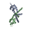
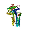
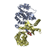
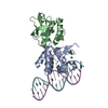
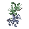
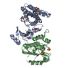
| Title | Dimeric Rous Sarcoma virus Capsid protein structure with an upstream 25-amino acid residue extension of C-terminal of Gag p10 protein | ||||||
 Components Components | GAG POLYPROTEIN CAPSID PROTEIN P27 | ||||||
 Keywords Keywords | VIRAL PROTEIN / Retrovirus / capsid protein / Gag Polyprotein / immature gag | ||||||
| Function / homology |  Function and homology information Function and homology informationhost cell nucleoplasm / viral procapsid maturation / host cell nucleolus / Hydrolases; Acting on peptide bonds (peptidases); Aspartic endopeptidases / viral capsid / structural constituent of virion / aspartic-type endopeptidase activity / nucleic acid binding / viral translational frameshifting / host cell plasma membrane ...host cell nucleoplasm / viral procapsid maturation / host cell nucleolus / Hydrolases; Acting on peptide bonds (peptidases); Aspartic endopeptidases / viral capsid / structural constituent of virion / aspartic-type endopeptidase activity / nucleic acid binding / viral translational frameshifting / host cell plasma membrane / proteolysis / zinc ion binding Similarity search - Function | ||||||
| Biological species |  Rous sarcoma virus Rous sarcoma virus | ||||||
| Method |  X-RAY DIFFRACTION / SYNCHROTRON / MOLECULAR REPLACEMENT / Resolution: 2.6 Å X-RAY DIFFRACTION / SYNCHROTRON / MOLECULAR REPLACEMENT / Resolution: 2.6 Å | ||||||
 Authors Authors | Nandhagopal, N. / Simpson, A.A. / Johnson, M.C. / Francisco, A.B. / Schatz, G.W. / Rossmann, M.G. / Vogt, V.M. | ||||||
 Citation Citation | Journal: J.Mol.Biol. / Year: 2004 Title: Dimeric rous sarcoma virus capsid protein structure relevant to immature gag assembly Authors: Nandhagopal, N. / Simpson, A.A. / Johnson, M.C. / Francisco, A.B. / Schatz, G.W. / Rossmann, M.G. / Vogt, V.M. | ||||||
| History |
| ||||||
| Remark 999 | SEQUENCE The protein sequence contains the N-terminal domain of capsid protein P27 with an upstream ...SEQUENCE The protein sequence contains the N-terminal domain of capsid protein P27 with an upstream 25-amino acid extension C-terminal of GAG P10 PROTEIN. |
- Structure visualization
| Structure viewer | Molecule: MolmilJmol/JSmol |
|---|
- Downloads & links
-Download
| PDBx/mmCIF format | 1p7n.cif.gz | 44.2 KB | Display | PDBx/mmCIF format |
|---|---|---|---|---|
| PDB format | pdb1p7n.ent.gz | 31.2 KB | Display | PDB format |
| PDBx/mmJSON format | 1p7n.json.gz | Tree view | PDBx/mmJSON format | |
| Others |  Other downloads Other downloads |
-Validation report
| Arichive directory | https://data.pdbj.org/pub/pdb/validation_reports/p7/1p7nftp://data.pdbj.org/pub/pdb/validation_reports/p7/1p7n | HTTPS FTP |
|---|
-Related structure data
| Related structure data |  1em9S S: Starting model for refinement |
|---|---|
| Similar structure data |


-Links
 PDBj
PDBj- Assembly
| Deposited unit | 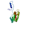
| ||||||||
|---|---|---|---|---|---|---|---|---|---|
| 1 | 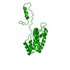
| ||||||||
| Unit cell |
|
-Components
| #1: Protein | Mass: 18736.674 Da / Num. of mol.: 1 / Fragment: N-terminal domain Source method: isolated from a genetically manipulated source Source: (gene. exp.) Rous sarcoma virus / Genus: Alpharetrovirus / Production host:  |
|---|
-Experimental details
-Experiment
| Experiment | Method: X-RAY DIFFRACTION / Number of used crystals: 1 |
|---|
- Sample preparation
| Crystal | Density Matthews: 2.6 Å3/Da / Density % sol: 52.73 % | ||||||||||||||||||||||||||||||||||||||||||
|---|---|---|---|---|---|---|---|---|---|---|---|---|---|---|---|---|---|---|---|---|---|---|---|---|---|---|---|---|---|---|---|---|---|---|---|---|---|---|---|---|---|---|---|
| Crystal grow | Temperature: 293 K / Method: vapor diffusion, hanging drop / pH: 7.5 Details: 10 mM HEPES-sodium buffer and 0.8M potassium sodium tartrate tetrahydrate, pH 7.5, VAPOR DIFFUSION, HANGING DROP, temperature 293K | ||||||||||||||||||||||||||||||||||||||||||
| Crystal grow | *PLUS Temperature: 20 ℃ / pH: 8 / Method: vapor diffusion | ||||||||||||||||||||||||||||||||||||||||||
| Components of the solutions | *PLUS
|
-Data collection
| Diffraction | Mean temperature: 298 K |
|---|---|
| Diffraction source | Source: SYNCHROTRON / Site: APS  / Beamline: 14-ID-B / Wavelength: 0.9 Å / Beamline: 14-ID-B / Wavelength: 0.9 Å |
| Detector | Type: ADSC QUANTUM 4 / Detector: CCD / Date: Jun 20, 2002 |
| Radiation | Protocol: SINGLE WAVELENGTH / Monochromatic (M) / Laue (L): M / Scattering type: x-ray |
| Radiation wavelength | Wavelength: 0.9 Å / Relative weight: 1 |
| Reflection | Resolution: 2.45→30 Å / Num. all: 7911 / Num. obs: 6743 / % possible obs: 85.2 % / Redundancy: 8.3 % / Biso Wilson estimate: 20.3 Å2 / Rmerge(I) obs: 0.055 |
| Reflection shell | Resolution: 2.45→2.54 Å / Rmerge(I) obs: 0.329 / Num. unique all: 396 / % possible all: 51.4 |
| Reflection | *PLUS Redundancy: 3.18 % |
| Reflection shell | *PLUS % possible obs: 51.4 % / Redundancy: 1.4 % |
- Processing
| Software |
| ||||||||||||||||||||||||||||||||||||
|---|---|---|---|---|---|---|---|---|---|---|---|---|---|---|---|---|---|---|---|---|---|---|---|---|---|---|---|---|---|---|---|---|---|---|---|---|---|
| Refinement | Method to determine structure: MOLECULAR REPLACEMENT Starting model: PDB ENTRY 1EM9 Resolution: 2.6→26.5 Å / Rfactor Rfree error: 0.016 / Data cutoff high absF: 558128.82 / Data cutoff high rms absF: 558128.82 / Data cutoff low absF: 0 / Isotropic thermal model: RESTRAINED / Cross valid method: THROUGHOUT / σ(F): 0
| ||||||||||||||||||||||||||||||||||||
| Solvent computation | Solvent model: FLAT MODEL / Bsol: 38.5603 Å2 / ksol: 0.303224 e/Å3 | ||||||||||||||||||||||||||||||||||||
| Displacement parameters | Biso mean: 56.4 Å2
| ||||||||||||||||||||||||||||||||||||
| Refine analyze |
| ||||||||||||||||||||||||||||||||||||
| Refinement step | Cycle: LAST / Resolution: 2.6→26.5 Å
| ||||||||||||||||||||||||||||||||||||
| Refine LS restraints |
| ||||||||||||||||||||||||||||||||||||
| LS refinement shell | Resolution: 2.6→2.76 Å / Rfactor Rfree error: 0.062 / Total num. of bins used: 6
| ||||||||||||||||||||||||||||||||||||
| Xplor file |
| ||||||||||||||||||||||||||||||||||||
| Refinement | *PLUS Highest resolution: 2.6 Å / % reflection Rfree: 5 % / Rfactor Rwork: 0.26 | ||||||||||||||||||||||||||||||||||||
| Solvent computation | *PLUS | ||||||||||||||||||||||||||||||||||||
| Displacement parameters | *PLUS | ||||||||||||||||||||||||||||||||||||
| Refine LS restraints | *PLUS
| ||||||||||||||||||||||||||||||||||||
| LS refinement shell | *PLUS Rfactor Rfree: 0.366 / Rfactor Rwork: 0.337 |
