 Movie
Movie Controller

[English] 日本語
 Yorodumi
- PDB-1p6l: Bovine endothelial NOS heme domain with L-N(omega)-nitroarginine-... -
+ Open data

- Basic information
| Entry | Database: PDB / ID: 1p6l | ||||||
|---|---|---|---|---|---|---|---|
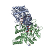
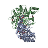
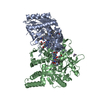
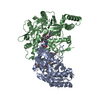
| Title | Bovine endothelial NOS heme domain with L-N(omega)-nitroarginine-2,4-L-diaminobutyric amide bound | ||||||
 Components Components | Nitric-oxide synthase, endothelial | ||||||
 Keywords Keywords | OXIDOREDUCTASE / nitric oxide synthase / heme-enzyme | ||||||
| Function / homology |  Function and homology information Function and homology informationcellular response to laminar fluid shear stress / negative regulation of leukocyte cell-cell adhesion / nitric-oxide synthase (NADPH) / : / nitric-oxide synthase activity / L-arginine catabolic process / negative regulation of extrinsic apoptotic signaling pathway via death domain receptors / nitric oxide biosynthetic process / negative regulation of blood pressure / response to hormone ...cellular response to laminar fluid shear stress / negative regulation of leukocyte cell-cell adhesion / nitric-oxide synthase (NADPH) / : / nitric-oxide synthase activity / L-arginine catabolic process / negative regulation of extrinsic apoptotic signaling pathway via death domain receptors / nitric oxide biosynthetic process / negative regulation of blood pressure / response to hormone / positive regulation of relaxation of smooth muscle / mitochondrion organization / caveola / response to peptide hormone / blood coagulation / NADP binding / FMN binding / flavin adenine dinucleotide binding / response to lipopolysaccharide / cytoskeleton / calmodulin binding / heme binding / Golgi apparatus / metal ion binding / nucleus / plasma membrane / cytosol Similarity search - Function | ||||||
| Biological species |  | ||||||
| Method |  X-RAY DIFFRACTION / SYNCHROTRON / FOURIER SYNTHESIS / Resolution: 2.35 Å X-RAY DIFFRACTION / SYNCHROTRON / FOURIER SYNTHESIS / Resolution: 2.35 Å | ||||||
 Authors Authors | Flinspach, M.L. / Li, H. / Jamal, J. / Yang, W. / Huang, H. / Hah, J.-M. / Gomez-Vidal, J.A. / Litzinger, E.A. / Silverman, R.B. / Poulos, T.L. | ||||||
 Citation Citation | Journal: Nat.Struct.Mol.Biol. / Year: 2004 Title: Structural basis for dipeptide amide isoform-selective inhibition of neuronal nitric oxide synthase. Authors: Flinspach, M.L. / Li, H. / Jamal, J. / Yang, W. / Huang, H. / Hah, J.M. / Gomez-Vidal, J.A. / Litzinger, E.A. / Silverman, R.B. / Poulos, T.L. #1: Journal: Cell(Cambridge,Mass.) / Year: 1998Title: CRYSTAL STRUCTURE OF CONSTITUTIVE ENDOTHELIAL NITRIC OXIDE SYNTHASE: A PARADIGM FOR PTERIN FUNCTION INVOLVING A NOVEL METAL CENTER Authors: Raman, C.S. / Li, H. / Martasek, P. / Kral, V. / Masters, B.S.S. / Poulos, T.L. #2: Journal: Biochemistry / Year: 2002Title: The novel binding mode of N-alkyl-N'-hydroxyguanidine to neuronal nitric oxide synthase provides mechanistic insights into NO biosynthesis Authors: Li, H. / Shimizu, H. / Flinspach, M. / Jamal, J. / Yang, W. / Xian, M. / Cai, T. / Wen, E.Z. / Jia, Q. / Wang, P.G. / Poulos, T.L. | ||||||
| History |
| ||||||
| Remark 999 | SEQUENCE AUTHOR STATED THAT STRUCTURAL DATA CLEARLY INDICATED THAT RESIDUE 100 IS ARG RATHER THAN CYS. |
- Structure visualization
| Structure viewer | Molecule: MolmilJmol/JSmol |
|---|
- Downloads & links
-Download
| PDBx/mmCIF format | 1p6l.cif.gz | 190 KB | Display | PDBx/mmCIF format |
|---|---|---|---|---|
| PDB format | pdb1p6l.ent.gz | 147.4 KB | Display | PDB format |
| PDBx/mmJSON format | 1p6l.json.gz | Tree view | PDBx/mmJSON format | |
| Others |  Other downloads Other downloads |
-Validation report
| Arichive directory | https://data.pdbj.org/pub/pdb/validation_reports/p6/1p6lftp://data.pdbj.org/pub/pdb/validation_reports/p6/1p6l | HTTPS FTP |
|---|
-Related structure data
| Related structure data |  1p6hC  1p6iC  1p6jC  1p6kC  1p6mC  1p6nC  1q2oC C: citing same article ( |
|---|---|
| Similar structure data |


-Links
 PDBj
PDBj


- Assembly
| Deposited unit | 
| ||||||||
|---|---|---|---|---|---|---|---|---|---|
| 1 |
| ||||||||
| Unit cell |
|
-Components
-Protein , 1 types, 2 molecules AB
| #1: Protein | Mass: 46983.207 Da / Num. of mol.: 2 / Fragment: NOS HEME DOMAIN Source method: isolated from a genetically manipulated source Source: (gene. exp.)  Pichia pastoris (fungus) / Strain (production host): KM71H / References: UniProt: P29473, nitric-oxide synthase (NADPH) Pichia pastoris (fungus) / Strain (production host): KM71H / References: UniProt: P29473, nitric-oxide synthase (NADPH) |
|---|
-Non-polymers , 8 types, 455 molecules 


| #2: Chemical |  Mass: 136.989 Da / Num. of mol.: 2 / Source method: obtained synthetically / Formula: C2H6AsO2 Mass: 136.989 Da / Num. of mol.: 2 / Source method: obtained synthetically / Formula: C2H6AsO2#3: Chemical |  Mass: 59.044 Da / Num. of mol.: 2 / Source method: obtained synthetically / Formula: C2H3O2 Mass: 59.044 Da / Num. of mol.: 2 / Source method: obtained synthetically / Formula: C2H3O2#4: Chemical |  Mass: 616.487 Da / Num. of mol.: 2 / Source method: obtained synthetically / Formula: C34H32FeN4O4 Mass: 616.487 Da / Num. of mol.: 2 / Source method: obtained synthetically / Formula: C34H32FeN4O4#5: Chemical |  Mass: 241.247 Da / Num. of mol.: 2 / Source method: obtained synthetically / Formula: C9H15N5O3 Mass: 241.247 Da / Num. of mol.: 2 / Source method: obtained synthetically / Formula: C9H15N5O3#6: Chemical |  Mass: 318.333 Da / Num. of mol.: 2 / Source method: obtained synthetically / Formula: C10H22N8O4 Mass: 318.333 Da / Num. of mol.: 2 / Source method: obtained synthetically / Formula: C10H22N8O4#7: Chemical |  Mass: 92.094 Da / Num. of mol.: 2 / Source method: obtained synthetically / Formula: C3H8O3 Mass: 92.094 Da / Num. of mol.: 2 / Source method: obtained synthetically / Formula: C3H8O3#8: Chemical | ChemComp-ZN / |  Mass: 65.409 Da / Num. of mol.: 1 / Source method: obtained synthetically / Formula: Zn Mass: 65.409 Da / Num. of mol.: 1 / Source method: obtained synthetically / Formula: Zn#9: Water | ChemComp-HOH / | Mass: 18.015 Da / Num. of mol.: 442 / Source method: isolated from a natural source / Formula: H2O |
|---|
-Experimental details
-Experiment
| Experiment | Method: X-RAY DIFFRACTION / Number of used crystals: 1 |
|---|
- Sample preparation
| Crystal | Density Matthews: 2.55 Å3/Da / Density % sol: 51.83 % | ||||||||||||||||||||||||||||||||||||||||||||||||||||||
|---|---|---|---|---|---|---|---|---|---|---|---|---|---|---|---|---|---|---|---|---|---|---|---|---|---|---|---|---|---|---|---|---|---|---|---|---|---|---|---|---|---|---|---|---|---|---|---|---|---|---|---|---|---|---|---|
| Crystal grow | Temperature: 280 K / Method: vapor diffusion, sitting drop / pH: 6.9 Details: PEG3350, MgOAc, Na Cacodylate, TCEP, pH 6.9, VAPOR DIFFUSION, SITTING DROP, temperature 280K | ||||||||||||||||||||||||||||||||||||||||||||||||||||||
| Crystal grow | *PLUS Temperature: 5 ℃ / Method: vapor diffusion, sitting drop / Details: Li, H., (2002) Biochemistry, 41, 13868. / PH range low: 6 / PH range high: 5.6 | ||||||||||||||||||||||||||||||||||||||||||||||||||||||
| Components of the solutions | *PLUS
|
-Data collection
| Diffraction | Mean temperature: 100 K |
|---|---|
| Diffraction source | Source: SYNCHROTRON / Site: SSRL  / Beamline: BL7-1 / Wavelength: 1.08 Å / Beamline: BL7-1 / Wavelength: 1.08 Å |
| Detector | Type: MARRESEARCH / Detector: IMAGE PLATE / Date: Nov 26, 2001 / Details: mirrors |
| Radiation | Monochromator: yes / Protocol: SINGLE WAVELENGTH / Monochromatic (M) / Laue (L): M / Scattering type: x-ray |
| Radiation wavelength | Wavelength: 1.08 Å / Relative weight: 1 |
| Reflection | Resolution: 2.35→50 Å / Num. obs: 37915 / % possible obs: 92.2 % / Observed criterion σ(I): -3 / Redundancy: 3.4 % / Biso Wilson estimate: 40.5 Å2 / Rmerge(I) obs: 0.084 / Rsym value: 0.084 / Net I/σ(I): 14.26 |
| Reflection shell | Resolution: 2.35→2.39 Å / Redundancy: 3 % / Rmerge(I) obs: 0.522 / Mean I/σ(I) obs: 1.9 / Rsym value: 0.522 / % possible all: 60.7 |
| Reflection | *PLUS Num. measured all: 126627 |
| Reflection shell | *PLUS % possible obs: 60.7 % / Rmerge(I) obs: 0.552 / Mean I/σ(I) obs: 2 |
- Processing
| Software |
| ||||||||||||||||||||
|---|---|---|---|---|---|---|---|---|---|---|---|---|---|---|---|---|---|---|---|---|---|
| Refinement | Method to determine structure: FOURIER SYNTHESIS / Resolution: 2.35→34.98 Å / Rfactor Rfree error: 0.006 / Isotropic thermal model: RESTRAINED / Cross valid method: THROUGHOUT / σ(F): 0 / Stereochemistry target values: Engh & Huber
| ||||||||||||||||||||
| Solvent computation | Solvent model: FLAT MODEL / Bsol: 35.6823 Å2 / ksol: 0.357765 e/Å3 | ||||||||||||||||||||
| Displacement parameters | Biso mean: 42.6 Å2
| ||||||||||||||||||||
| Refine analyze |
| ||||||||||||||||||||
| Refinement step | Cycle: LAST / Resolution: 2.35→34.98 Å
| ||||||||||||||||||||
| Refine LS restraints |
| ||||||||||||||||||||
| LS refinement shell | Resolution: 2.35→2.43 Å / Rfactor Rfree error: 0.032 / Total num. of bins used: 10
| ||||||||||||||||||||
| Xplor file |
| ||||||||||||||||||||
| Refinement | *PLUS % reflection Rfree: 5 % | ||||||||||||||||||||
| Solvent computation | *PLUS | ||||||||||||||||||||
| Displacement parameters | *PLUS | ||||||||||||||||||||
| Refine LS restraints | *PLUS
|
