 Movie
Movie Controller

[English] 日本語
 Yorodumi
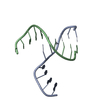
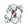
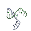
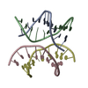
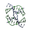
- PDB-1p54: Effect of Sequence on the Conformational Geometry of DNA Holliday... -
+ Open data

- Basic information
| Entry | Database: PDB / ID: 1p54 | ||||||||||||||||||
|---|---|---|---|---|---|---|---|---|---|---|---|---|---|---|---|---|---|---|---|
| Title | Effect of Sequence on the Conformational Geometry of DNA Holliday Junctions | ||||||||||||||||||
 Components Components | 5'-D(* Keywords KeywordsDNA / DNA Holliday Junction / Four-Way Junction / stacked-X junction / brominated uracil | Function / homology | DNA |  Function and homology information Function and homology informationMethod |  X-RAY DIFFRACTION / MOLECULAR REPLACEMENT / Resolution: 1.9 Å X-RAY DIFFRACTION / MOLECULAR REPLACEMENT / Resolution: 1.9 Å  Authors AuthorsHays, F.A. / Vargason, J.M. / Ho, P.S. |  CitationJournal: Biochemistry / Year: 2003 CitationJournal: Biochemistry / Year: 2003Title: Effect of Sequence on the Conformation of DNA Holliday Junctions Authors: Hays, F.A. / Vargason, J.M. / Ho, P.S. History |
|
- Structure visualization
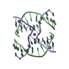
| Structure viewer | Molecule: MolmilJmol/JSmol |
|---|
- Downloads & links
-Download
| PDBx/mmCIF format | 1p54.cif.gz | 23.4 KB | Display | PDBx/mmCIF format |
|---|---|---|---|---|
| PDB format | pdb1p54.ent.gz | 14.9 KB | Display | PDB format |
| PDBx/mmJSON format | 1p54.json.gz | Tree view | PDBx/mmJSON format | |
| Others |  Other downloads Other downloads |
-Validation report
| Arichive directory | https://data.pdbj.org/pub/pdb/validation_reports/p5/1p54ftp://data.pdbj.org/pub/pdb/validation_reports/p5/1p54 | HTTPS FTP |
|---|
-Related structure data
| Related structure data |  1p4yC 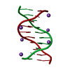 1p4zC 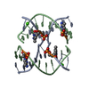 1l6bS C: citing same article ( S: Starting model for refinement |
|---|---|
| Similar structure data |

-Links
 PDBj
PDBj

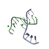
- Assembly
| Deposited unit | 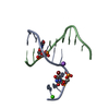
| ||||||||||||
|---|---|---|---|---|---|---|---|---|---|---|---|---|---|
| 1 | 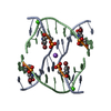
| ||||||||||||
| Unit cell |
| ||||||||||||
| Components on special symmetry positions |
| ||||||||||||
| Details | This entry contains the crystallographic asymmetric unit which consists of two chains A and B. The full four stranded Holliday junction structure can be generated by the following: matrix = (-1.00000 0.00001 0.00000) (0.00001 1.00000 -0.00002) (0.00000 -0.00002 -1.00000) translation = (0.00022 0.00018 -0.00034) |
-Components
| #1: DNA chain | Mass: 3109.874 Da / Num. of mol.: 2 / Source method: obtained synthetically Details: DNA was synthesized on an Applied Biosystems DNA synthesizer using phosphoramidite chemistry, with the trityl-protecting group left intact at the 5-terminal nucleotide, FOR SUBSEQUENT HPLC ...Details: DNA was synthesized on an Applied Biosystems DNA synthesizer using phosphoramidite chemistry, with the trityl-protecting group left intact at the 5-terminal nucleotide, FOR SUBSEQUENT HPLC PURIFICATION, THEN DEPROTECTED BY TREATMENT WITH 3% ACETIC ACID FOR FIFTEEN MINUTES, NEUTRALIZED WITH AMMONIUM HYDROXIDE, AND DESALTED ON A SIGMA G-25 SEPHADEX COLUMN. #2: Chemical | ChemComp-NA / |   Mass: 22.990 Da / Num. of mol.: 1 / Source method: obtained synthetically / Formula: Na Mass: 22.990 Da / Num. of mol.: 1 / Source method: obtained synthetically / Formula: Na#3: Chemical | ChemComp-CA / |   Mass: 40.078 Da / Num. of mol.: 1 / Source method: obtained synthetically / Formula: Ca Mass: 40.078 Da / Num. of mol.: 1 / Source method: obtained synthetically / Formula: Ca#4: Water | ChemComp-HOH / |  Mass: 18.015 Da / Num. of mol.: 87 / Source method: isolated from a natural source / Formula: H2O Mass: 18.015 Da / Num. of mol.: 87 / Source method: isolated from a natural source / Formula: H2O |
|---|
-Experimental details
-Experiment
| Experiment | Method: X-RAY DIFFRACTION / Number of used crystals: 1 |
|---|
- Sample preparation
| Crystal | Density Matthews: 1.93 Å3/Da / Density % sol: 35.87 % | ||||||||||||||||||||||||||||||||||||||||||||||||||||||
|---|---|---|---|---|---|---|---|---|---|---|---|---|---|---|---|---|---|---|---|---|---|---|---|---|---|---|---|---|---|---|---|---|---|---|---|---|---|---|---|---|---|---|---|---|---|---|---|---|---|---|---|---|---|---|---|
| Crystal grow | Temperature: 298 K / pH: 7.5 Details: 0.6mM DNA, 5mM Tris-HCL, 120mM calcium acetate, 16% 2-methyl-2,4-pentane diol, pH 7.5, VAPOR DIFFUSION, SITTING DROP, temperature 298K, pH 7.50 | ||||||||||||||||||||||||||||||||||||||||||||||||||||||
| Components of the solutions |
| ||||||||||||||||||||||||||||||||||||||||||||||||||||||
| Crystal grow | *PLUS pH: 7.5 / Method: vapor diffusion | ||||||||||||||||||||||||||||||||||||||||||||||||||||||
| Components of the solutions | *PLUS
|
-Data collection
| Diffraction | Mean temperature: 103 K |
|---|---|
| Diffraction source | Source: ROTATING ANODE / Type: RIGAKU RU300 / Wavelength: 1.542 |
| Detector | Type: RIGAKU RAXIS IV / Detector: IMAGE PLATE / Date: Dec 19, 2002 |
| Radiation | Monochromator: NI FILTER / Protocol: SINGLE WAVELENGTH / Monochromatic (M) / Laue (L): M / Scattering type: x-ray |
| Radiation wavelength | Wavelength: 1.542 Å / Relative weight: 1 |
| Reflection | Resolution: 1.8→19.18 Å / Num. obs: 4177 / % possible obs: 82 % / Observed criterion σ(I): 0 / Biso Wilson estimate: 4.3 Å2 / Rmerge(I) obs: 0.041 / Net I/σ(I): 4.05 |
| Reflection shell | Resolution: 1.8→1.86 Å / Rmerge(I) obs: 0.189 / % possible all: 34.9 |
| Reflection | *PLUS Highest resolution: 1.8 Å / Lowest resolution: 50 Å / Num. obs: 4011 / Num. measured all: 13013 |
| Reflection shell | *PLUS % possible obs: 34.9 % / Mean I/σ(I) obs: 4.1 |
- Processing
| Software |
| ||||||||||||||||||||||||||||||||||||||||||||||||||||||||||||
|---|---|---|---|---|---|---|---|---|---|---|---|---|---|---|---|---|---|---|---|---|---|---|---|---|---|---|---|---|---|---|---|---|---|---|---|---|---|---|---|---|---|---|---|---|---|---|---|---|---|---|---|---|---|---|---|---|---|---|---|---|---|
| Refinement | Method to determine structure: MOLECULAR REPLACEMENT Starting model: 1L6B USED FOR INITIAL SOLUTION VIA MOLECULAR REPLACEMENT Resolution: 1.9→19.18 Å / Rfactor Rfree error: 0.012 / Isotropic thermal model: RESTRAINED / Cross valid method: THROUGHOUT / σ(F): 0
| ||||||||||||||||||||||||||||||||||||||||||||||||||||||||||||
| Displacement parameters | Biso mean: 9.5 Å2 | ||||||||||||||||||||||||||||||||||||||||||||||||||||||||||||
| Refine analyze |
| ||||||||||||||||||||||||||||||||||||||||||||||||||||||||||||
| Refinement step | Cycle: LAST / Resolution: 1.9→19.18 Å
| ||||||||||||||||||||||||||||||||||||||||||||||||||||||||||||
| Refine LS restraints |
| ||||||||||||||||||||||||||||||||||||||||||||||||||||||||||||
| LS refinement shell | Resolution: 1.9→2.02 Å / Rfactor Rfree error: 0.041 / Total num. of bins used: 6
| ||||||||||||||||||||||||||||||||||||||||||||||||||||||||||||
| Refinement | *PLUS Highest resolution: 1.9 Å / Lowest resolution: 20 Å / % reflection Rfree: 10 % / Rfactor Rfree: 0.243 | ||||||||||||||||||||||||||||||||||||||||||||||||||||||||||||
| Solvent computation | *PLUS | ||||||||||||||||||||||||||||||||||||||||||||||||||||||||||||
| Displacement parameters | *PLUS | ||||||||||||||||||||||||||||||||||||||||||||||||||||||||||||
| Refine LS restraints | *PLUS
|
