 Movie
Movie Controller

[English] 日本語
 Yorodumi
- PDB-1ozv: Crystal structure of the SET domain of LSMT bound to Lysine and AdoHcy -
+ Open data

- Basic information
| Entry | Database: PDB / ID: 1ozv | ||||||
|---|---|---|---|---|---|---|---|
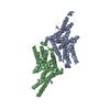
| Title | Crystal structure of the SET domain of LSMT bound to Lysine and AdoHcy | ||||||
 Components Components | Ribulose-1,5 bisphosphate carboxylase/oxygenase large subunit N-methyltransferase, chloroplast | ||||||
 Keywords Keywords | TRANSFERASE / SET DOMAIN / LYSINE N-METHYLATION / MULTIPLE METHYLATION / PHOTOSYNTHESIS / POST-TRANSLATIONAL MODIFICATION | ||||||
| Function / homology |  Function and homology information Function and homology information[ribulose-bisphosphate carboxylase]-lysine N-methyltransferase / [fructose-bisphosphate aldolase]-lysine N-methyltransferase / [ribulose-bisphosphate carboxylase]-lysine N-methyltransferase activity / protein-lysine N-methyltransferase activity / chloroplast / methylation Similarity search - Function | ||||||
| Biological species |  Pisum sativum (garden pea) Pisum sativum (garden pea) | ||||||
| Method |  X-RAY DIFFRACTION / MOLECULAR REPLACEMENT / Resolution: 2.65 Å X-RAY DIFFRACTION / MOLECULAR REPLACEMENT / Resolution: 2.65 Å | ||||||
 Authors Authors | Trievel, R.C. / Flynn, E.M. / Houtz, R.L. / Hurley, J.H. | ||||||
 Citation Citation | Journal: Nat.Struct.Biol. / Year: 2003 Title: Mechanism of multiple lysine methylation by the SET domain enzyme Rubisco LSMT Authors: Trievel, R.C. / Flynn, E.M. / Houtz, R.L. / Hurley, J.H. | ||||||
| History |
| ||||||
| Remark 999 | SEQUENCE AUTHORS INFORMED THAT RESIDUES 483-488 RESULT FROM A C-TERMINAL TEV PROTEASE CLEAVAGE SITE ...SEQUENCE AUTHORS INFORMED THAT RESIDUES 483-488 RESULT FROM A C-TERMINAL TEV PROTEASE CLEAVAGE SITE THAT WAS ENGINEERED INTO THE ENZYME AFTER LEU 482. AFTER CLEAVAGE, THESE SIX RESIDUES WERE LEFT ON THE C-TERMINUS OF THIS CONSTRUCT. |
- Structure visualization
| Structure viewer | Molecule: MolmilJmol/JSmol |
|---|
- Downloads & links
-Download
| PDBx/mmCIF format | 1ozv.cif.gz | 286.8 KB | Display | PDBx/mmCIF format |
|---|---|---|---|---|
| PDB format | pdb1ozv.ent.gz | 230.4 KB | Display | PDB format |
| PDBx/mmJSON format | 1ozv.json.gz | Tree view | PDBx/mmJSON format | |
| Others |  Other downloads Other downloads |
-Validation report
| Arichive directory | https://data.pdbj.org/pub/pdb/validation_reports/oz/1ozvftp://data.pdbj.org/pub/pdb/validation_reports/oz/1ozv | HTTPS FTP |
|---|
-Related structure data
| Related structure data |  1p0yC  1mlvS S: Starting model for refinement C: citing same article ( |
|---|---|
| Similar structure data |


-Links
 PDBj
PDBj- Assembly
| Deposited unit | 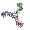
| ||||||||
|---|---|---|---|---|---|---|---|---|---|
| 1 | 
| ||||||||
| 2 | 
| ||||||||
| 3 | 
| ||||||||
| 4 |
| ||||||||
| Unit cell |
|
-Components
| #1: Protein | Mass: 50629.160 Da / Num. of mol.: 3 Source method: isolated from a genetically manipulated source Source: (gene. exp.) Pisum sativum (garden pea) / Plasmid: pDEST14 / Species (production host): Escherichia coli / Production host:  References: UniProt: Q43088, [ribulose-bisphosphate carboxylase]-lysine N-methyltransferase #2: Chemical |   Type: L-peptide linking / Mass: 147.195 Da / Num. of mol.: 3 / Source method: obtained synthetically / Formula: C6H15N2O2 Type: L-peptide linking / Mass: 147.195 Da / Num. of mol.: 3 / Source method: obtained synthetically / Formula: C6H15N2O2#3: Chemical |   Type: L-peptide linking / Mass: 384.411 Da / Num. of mol.: 3 / Source method: obtained synthetically / Formula: C14H20N6O5S Type: L-peptide linking / Mass: 384.411 Da / Num. of mol.: 3 / Source method: obtained synthetically / Formula: C14H20N6O5S#4: Water | ChemComp-HOH / |  Mass: 18.015 Da / Num. of mol.: 644 / Source method: isolated from a natural source / Formula: H2O Mass: 18.015 Da / Num. of mol.: 644 / Source method: isolated from a natural source / Formula: H2O |
|---|
-Experimental details
-Experiment
| Experiment | Method: X-RAY DIFFRACTION / Number of used crystals: 1 |
|---|
- Sample preparation
| Crystal | Density Matthews: 4.38 Å3/Da / Density % sol: 71.67 % | ||||||||||||||||||||||||||||||||||||
|---|---|---|---|---|---|---|---|---|---|---|---|---|---|---|---|---|---|---|---|---|---|---|---|---|---|---|---|---|---|---|---|---|---|---|---|---|---|
| Crystal grow | Temperature: 298 K / Method: vapor diffusion, hanging drop / pH: 6.8 Details: 0.95-1.10 M Sodium Acetate, 100 mM Lysine Acetate, 1 mM TCEP, 400 uM S-adenosylhomocysteine, pH 6.8, VAPOR DIFFUSION, HANGING DROP, temperature 298.0K | ||||||||||||||||||||||||||||||||||||
| Crystal grow | *PLUS Temperature: 25 ℃ / Method: vapor diffusion, hanging drop | ||||||||||||||||||||||||||||||||||||
| Components of the solutions | *PLUS
|
-Data collection
| Diffraction | Mean temperature: 95 K |
|---|---|
| Diffraction source | Source: ROTATING ANODE / Type: RIGAKU RU200 / Wavelength: 1.5418 Å |
| Detector | Type: RIGAKU RAXIS IV / Detector: IMAGE PLATE / Date: Dec 2, 2002 / Details: Osmic Confocal Mirrors |
| Radiation | Protocol: SINGLE WAVELENGTH / Monochromatic (M) / Laue (L): M / Scattering type: x-ray |
| Radiation wavelength | Wavelength: 1.5418 Å / Relative weight: 1 |
| Reflection | Resolution: 2.65→30 Å / Num. all: 80333 / Num. obs: 79741 / % possible obs: 98.9 % / Observed criterion σ(F): -3 / Observed criterion σ(I): -3 / Redundancy: 4.2 % / Biso Wilson estimate: 39.3 Å2 / Rsym value: 0.056 / Net I/σ(I): 25.9 |
| Reflection shell | Resolution: 2.65→2.74 Å / Mean I/σ(I) obs: 2.51 / Num. unique all: 7902 / Rsym value: 0.494 / % possible all: 99 |
| Reflection | *PLUS Rmerge(I) obs: 0.056 |
| Reflection shell | *PLUS % possible obs: 99 % / Num. unique obs: 7902 / Rmerge(I) obs: 0.494 |
- Processing
| Software |
| ||||||||||||||||||||||||||||||||||||
|---|---|---|---|---|---|---|---|---|---|---|---|---|---|---|---|---|---|---|---|---|---|---|---|---|---|---|---|---|---|---|---|---|---|---|---|---|---|
| Refinement | Method to determine structure: MOLECULAR REPLACEMENT Starting model: PDB Entry 1MLV Resolution: 2.65→29.76 Å / Rfactor Rfree error: 0.004 / Data cutoff high absF: 612033.97 / Data cutoff high rms absF: 612033.97 / Data cutoff low absF: 0 / Isotropic thermal model: RESTRAINED / Cross valid method: THROUGHOUT / σ(F): 0 / σ(I): 2.51 / Stereochemistry target values: Engh & Huber
| ||||||||||||||||||||||||||||||||||||
| Solvent computation | Solvent model: FLAT MODEL / Bsol: 49.2167 Å2 / ksol: 0.339194 e/Å3 | ||||||||||||||||||||||||||||||||||||
| Displacement parameters | Biso mean: 67.6 Å2
| ||||||||||||||||||||||||||||||||||||
| Refine analyze |
| ||||||||||||||||||||||||||||||||||||
| Refinement step | Cycle: LAST / Resolution: 2.65→29.76 Å
| ||||||||||||||||||||||||||||||||||||
| Refine LS restraints |
| ||||||||||||||||||||||||||||||||||||
| Refine LS restraints NCS | NCS model details: CONSTR | ||||||||||||||||||||||||||||||||||||
| LS refinement shell | Resolution: 2.65→2.82 Å / Rfactor Rfree error: 0.018 / Total num. of bins used: 6
| ||||||||||||||||||||||||||||||||||||
| Xplor file |
| ||||||||||||||||||||||||||||||||||||
| Refinement | *PLUS Lowest resolution: 30 Å / % reflection Rfree: 5 % | ||||||||||||||||||||||||||||||||||||
| Solvent computation | *PLUS | ||||||||||||||||||||||||||||||||||||
| Displacement parameters | *PLUS | ||||||||||||||||||||||||||||||||||||
| Refine LS restraints | *PLUS
|
