 Movie
Movie Controller

[English] 日本語
 Yorodumi
- PDB-1oys: Crystal Structure of the Phosphorolytic Exoribonuclease RNase PH ... -
+ Open data

- Basic information
| Entry | Database: PDB / ID: 1oys | ||||||
|---|---|---|---|---|---|---|---|
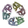
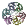
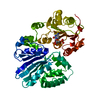
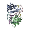
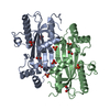
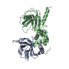
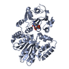
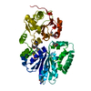
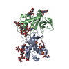
| Title | Crystal Structure of the Phosphorolytic Exoribonuclease RNase PH from Bacillus subtilis | ||||||
 Components Components | Ribonuclease PH | ||||||
 Keywords Keywords | TRANSFERASE / tRNA processing | ||||||
| Function / homology |  Function and homology information Function and homology informationtRNA nucleotidyltransferase / tRNA nucleotidyltransferase activity / rRNA 3'-end processing / rRNA catabolic process / tRNA processing / 3'-5'-RNA exonuclease activity / tRNA binding / RNA binding Similarity search - Function | ||||||
| Biological species |  | ||||||
| Method |  X-RAY DIFFRACTION / MOLECULAR REPLACEMENT / Resolution: 2.4 Å X-RAY DIFFRACTION / MOLECULAR REPLACEMENT / Resolution: 2.4 Å | ||||||
 Authors Authors | Harlow, L.S. / Kadziola, A. / Jensen, K.F. / Larsen, S. | ||||||
 Citation Citation | Journal: Protein Sci. / Year: 2004 Title: Crystal structure of the phosphorolytic exoribonuclease RNase PH from Bacillus subtilis and implications for its quaternary structure and tRNA binding. Authors: Harlow, L.S. / Kadziola, A. / Jensen, K.F. / Larsen, S. | ||||||
| History |
|
- Structure visualization
| Structure viewer | Molecule: MolmilJmol/JSmol |
|---|
- Downloads & links
-Download
| PDBx/mmCIF format | 1oys.cif.gz | 49.8 KB | Display | PDBx/mmCIF format |
|---|---|---|---|---|
| PDB format | pdb1oys.ent.gz | 36.7 KB | Display | PDB format |
| PDBx/mmJSON format | 1oys.json.gz | Tree view | PDBx/mmJSON format | |
| Others |  Other downloads Other downloads |
-Validation report
| Summary document | 1oys_validation.pdf.gz | 423.1 KB | Display | wwPDB validaton report |
|---|---|---|---|---|
| Full document | 1oys_full_validation.pdf.gz | 426.6 KB | Display | |
| Data in XML | 1oys_validation.xml.gz | 9.7 KB | Display | |
| Data in CIF | 1oys_validation.cif.gz | 12 KB | Display | |
| Arichive directory | https://data.pdbj.org/pub/pdb/validation_reports/oy/1oysftp://data.pdbj.org/pub/pdb/validation_reports/oy/1oys | HTTPS FTP |
-Related structure data


-Links
 PDBj
PDBj
- Assembly
| Deposited unit | 
| ||||||||
|---|---|---|---|---|---|---|---|---|---|
| 1 | 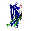
| ||||||||
| Unit cell |
|
-Components
| #1: Protein | Mass: 26626.244 Da / Num. of mol.: 1 / Mutation: R68Q,R73Q,R76Q Source method: isolated from a genetically manipulated source Source: (gene. exp.) |
|---|
-Experimental details
-Experiment
| Experiment | Method: X-RAY DIFFRACTION |
|---|
- Sample preparation
| Crystal | Density Matthews: 4.47 Å3/Da / Density % sol: 72.29 % | ||||||||||||||||||||||||
|---|---|---|---|---|---|---|---|---|---|---|---|---|---|---|---|---|---|---|---|---|---|---|---|---|---|
| Crystal grow | *PLUS Temperature: 20 ℃ / pH: 6.2 / Method: vapor diffusion, sitting drop | ||||||||||||||||||||||||
| Components of the solutions | *PLUS
|
-Data collection
| Radiation | Protocol: SINGLE WAVELENGTH / Monochromatic (M) / Laue (L): M / Scattering type: x-ray |
|---|---|
| Radiation wavelength | Relative weight: 1 |
| Reflection | Resolution: 2.4→20 Å / Num. obs: 16768 |
| Reflection | *PLUS Lowest resolution: 20 Å / % possible obs: 98.8 % / Redundancy: 4.4 % / Rmerge(I) obs: 0.047 |
| Reflection shell | *PLUS Highest resolution: 2.4 Å / Lowest resolution: 2.44 Å / % possible obs: 100 % / Redundancy: 5.5 % / Num. unique obs: 817 / Rmerge(I) obs: 0.206 / Mean I/σ(I) obs: 7 |
- Processing
| Software |
| ||||||||||||||||||||
|---|---|---|---|---|---|---|---|---|---|---|---|---|---|---|---|---|---|---|---|---|---|
| Refinement | Method to determine structure: MOLECULAR REPLACEMENT / Resolution: 2.4→20 Å / σ(F): 0
| ||||||||||||||||||||
| Refinement step | Cycle: LAST / Resolution: 2.4→20 Å
| ||||||||||||||||||||
| Refine LS restraints |
| ||||||||||||||||||||
| Refinement | *PLUS Lowest resolution: 20 Å / % reflection Rfree: 5 % / Rfactor Rfree: 0.284 / Rfactor Rwork: 0.258 | ||||||||||||||||||||
| Solvent computation | *PLUS | ||||||||||||||||||||
| Displacement parameters | *PLUS |
