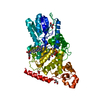
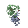
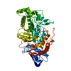
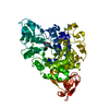
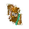
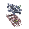
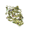
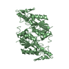
BIOMOLECULE: THIS ENTRY CONTAINS THE CRYSTALLOGRAPHIC ASYMMETRIC UNIT WHICH CONSISTS OF 3 CHAIN(S). ...BIOMOLECULE: THIS ENTRY CONTAINS THE CRYSTALLOGRAPHIC ASYMMETRIC UNIT WHICH CONSISTS OF 3 CHAIN(S). THE AUTHORS STATE THAT THE BIOLOGICAL UNIT IS NOT CLEAR.
Remark 400
COMPOUND THE AUTHORS STATE THE FOLLOWING: BASED ON THE PROTEIN SEQUENCE, THIS PROTEIN, AQ_TRMD, IS ...COMPOUND THE AUTHORS STATE THE FOLLOWING: BASED ON THE PROTEIN SEQUENCE, THIS PROTEIN, AQ_TRMD, IS ANNOTATED AS A TRNA (M1G37) METHYLTRANSFERASE, SAME AS ANOTHER TRNA (M1G37) METHYLTRANSFERASE OF KNOWN STRUCTURE (SUCH AS 1UAJ, 1UAK, 1UAL, 1UAM). THE MAJOR DIFFERENCE IS THAT THE LATTER STRUCTURES HAVE "TRIFOIL KNOTS", WHICH IS PROPOSED AS ESSENTIAL FOR S-ADENOSYL-L-METHIONINE BINDING, WHILE THIS STRUCTURE DOES NOT HAVE THE KNOT. THEREFORE, IT IS POSSIBLE THAT THE PROTEIN IN THIS ENTRY MAY HAVE A BIOCHEMICAL FUNCTION DIFFERENT FROM TRNA (1MG37) METHYLTRANSFERASE DESPITE THE SEQUENCE-BASED ANNOTATION AS SUCH.
Method to determine structure: MAD / Resolution: 2.6→20 Å / SU B: 20.353 / SU ML: 0.454 / TLS residual ADP flag: LIKELY RESIDUAL / Cross valid method: THROUGHOUT / ESU R: 1.086 / ESU R Free: 0.377 / Stereochemistry target values: MAXIMUM LIKELIHOOD / Details: HYDROGENS HAVE BEEN ADDED IN THE RIDING POSITIONS
Rfactor
Num. reflection
% reflection
Selection details
Rfree
0.30029
1312
5.2 %
RANDOM
Rwork
0.27937
-
-
-
obs
0.28048
24034
99.35 %
-
all
-
24191
-
-
Solvent computation
Ion probe radii: 0.8 Å / Shrinkage radii: 0.8 Å / VDW probe radii: 1.4 Å / Solvent model: BABINET MODEL WITH MASK
Displacement parameters
Biso mean: 29.025 Å2
Baniso -1
Baniso -2
Baniso -3
1-
-0.75 Å2
0 Å2
0.16 Å2
2-
-
1.89 Å2
0 Å2
3-
-
-
-1.1 Å2
Refinement step
Cycle: LAST / Resolution: 2.6→20 Å
Protein
Nucleic acid
Ligand
Solvent
Total
Num. atoms
5303
0
0
76
5379
LS refinement shell
Resolution: 2.6→2.666 Å / Total num. of bins used: 20 /
Rfactor
Num. reflection
Rfree
0.343
90
Rwork
0.298
1734
Refinement TLS params.
Method: refined / Refine-ID: X-RAY DIFFRACTION
ID
L11 (°2)
L12 (°2)
L13 (°2)
L22 (°2)
L23 (°2)
L33 (°2)
S11 (Å °)
S12 (Å °)
S13 (Å °)
S21 (Å °)
S22 (Å °)
S23 (Å °)
S31 (Å °)
S32 (Å °)
S33 (Å °)
T11 (Å2)
T12 (Å2)
T13 (Å2)
T22 (Å2)
T23 (Å2)
T33 (Å2)
Origin x (Å)
Origin y (Å)
Origin z (Å)
1
2.5478
0.5433
-0.6714
4.4106
0.2262
4.9878
-0.2551
-0.0388
-0.1883
0.0163
0.2507
-0.3595
0.2735
0.3899
0.0044
0.0666
0.0124
-0.0099
0.1279
-0.0875
0.1293
-37.5218
-10.192
9.4359
2
4.1335
-0.6957
0.8279
1.505
0.1464
3.927
0.0042
-0.096
0.0696
-0.0264
-0.0145
0.001
-0.0095
0.0265
0.0103
0.055
-0.0103
0.0013
0.0545
0.0043
0.0294
-14.3445
42.2779
24.2227
3
4.1294
2.1136
-1.3969
5.7814
-1.0157
3.6675
-0.0354
-0.2038
0.0383
-0.2687
-0.0343
-0.5903
-0.0397
0.1815
0.0696
0.1551
-0.0905
0.001
0.1425
-0.1462
0.2362
-25.3947
10.2309
4.8704
4
13.2934
0.5164
2.2644
6.5572
-2.5674
9.1935
-0.2886
0.5279
-2.4543
-1.2192
-0.3898
-1.4608
1.8869
1.0217
0.6784
1.0256
-0.166
0.7649
0.552
-0.1537
1.3512
-9.614
5.791
-9.9538
5
8.8721
-0.8664
3.2591
9.6893
-1.3787
3.1473
0.1145
-0.5782
1.6154
0.4607
-0.2011
0.9953
-0.9943
0.3086
0.0867
0.4929
-0.2334
0.1856
0.3624
-0.2241
0.5112
8.5896
57.6801
48.8469
6
18.5065
-3.7492
-4.2862
4.4409
4.2074
-1.0641
-0.813
-0.7501
0.3196
0.9449
0.9271
-1.0207
1.3593
0.4979
-0.1141
1.1212
0.5794
-0.3177
0.5548
0.0003
0.53
-26.7051
-20.3982
25.9528
Refinement TLS group
ID
Refine-ID
Refine TLS-ID
Auth asym-ID
Label asym-ID
Auth seq-ID
Label seq-ID
1
X-RAY DIFFRACTION
1
A
A
4 - 164
4 - 164
2
X-RAY DIFFRACTION
2
B
B
304 - 464
4 - 164
3
X-RAY DIFFRACTION
3
C
C
604 - 764
4 - 164
4
X-RAY DIFFRACTION
4
A
A
179 - 235
179 - 235
5
X-RAY DIFFRACTION
5
B
B
479 - 535
179 - 235
6
X-RAY DIFFRACTION
6
C
C
779 - 835
179 - 235
Refinement
*PLUS
Highest resolution: 2.6 Å / Lowest resolution: 20 Å / Rfactor Rfree: 0.3 / Rfactor Rwork: 0.279
Solvent computation
*PLUS
Displacement parameters
*PLUS
Refine LS restraints
*PLUS
Refine-ID
Type
Dev ideal
X-RAY DIFFRACTION
bond_d
0.018
X-RAY DIFFRACTION
angle_d
X-RAY DIFFRACTION
angle_deg
1.97
+
About Yorodumi
-
News
-
Feb 9, 2022. New format data for meta-information of EMDB entries
New format data for meta-information of EMDB entries
Version 3 of the EMDB header file is now the official format.
The previous official version 1.9 will be removed from the archive.
In the structure databanks used in Yorodumi, some data are registered as the other names, "COVID-19 virus" and "2019-nCoV". Here are the details of the virus and the list of structure data.
Jan 31, 2019. EMDB accession codes are about to change! (news from PDBe EMDB page)
EMDB accession codes are about to change! (news from PDBe EMDB page)
The allocation of 4 digits for EMDB accession codes will soon come to an end. Whilst these codes will remain in use, new EMDB accession codes will include an additional digit and will expand incrementally as the available range of codes is exhausted. The current 4-digit format prefixed with “EMD-” (i.e. EMD-XXXX) will advance to a 5-digit format (i.e. EMD-XXXXX), and so on. It is currently estimated that the 4-digit codes will be depleted around Spring 2019, at which point the 5-digit format will come into force.
The EM Navigator/Yorodumi systems omit the EMD- prefix.
Related info.:Q: What is EMD? / ID/Accession-code notation in Yorodumi/EM Navigator
Yorodumi is a browser for structure data from EMDB, PDB, SASBDB, etc.
This page is also the successor to EM Navigator detail page, and also detail information page/front-end page for Omokage search.
The word "yorodu" (or yorozu) is an old Japanese word meaning "ten thousand". "mi" (miru) is to see.
Related info.:EMDB / PDB / SASBDB / Comparison of 3 databanks / Yorodumi Search / Aug 31, 2016. New EM Navigator & Yorodumi / Yorodumi Papers / Jmol/JSmol / Function and homology information / Changes in new EM Navigator and Yorodumi
 Movie
Movie Controller

 Yorodumi
 Open data
 Basic information
 Components
 Keywords
 Function and homology information
 Aquifex aeolicus (bacteria)
 X-RAY DIFFRACTION /
  Authors
 Citation
 Structure visualization
 Downloads & links
 Other downloads
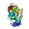

 PDBj
PDBj Assembly

 Mass: 18.015 Da / Num. of mol.: 76 / Source method: isolated from a natural source / Formula: H2O
 Sample preparation
 / Beamline: 5.0.2 / Wavelength: 0.9640,0.9793
 Processing