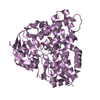
 Movie
Movie Controller


+ Open data

- Basic information
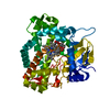
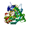
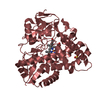
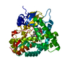
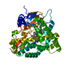
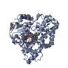
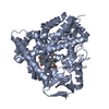
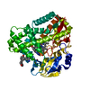
| Entry | Database: PDB / ID: 1oxa | ||||||
|---|---|---|---|---|---|---|---|
| Title | CYTOCHROME P450 (DONOR:O2 OXIDOREDUCTASE) | ||||||
 Components Components | CYTOCHROME P450 ERYF | ||||||
 Keywords Keywords | OXIDOREDUCTASE (OXYGENASE) | ||||||
| Function / homology |  Function and homology information Function and homology information6-deoxyerythronolide B hydroxylase / erythromycin biosynthetic process / oxidoreductase activity, acting on paired donors, with incorporation or reduction of molecular oxygen / monooxygenase activity / iron ion binding / heme binding / cytoplasm Similarity search - Function | ||||||
| Biological species |  Saccharopolyspora erythraea (bacteria) Saccharopolyspora erythraea (bacteria) | ||||||
| Method |  X-RAY DIFFRACTION / Resolution: 2.1 Å X-RAY DIFFRACTION / Resolution: 2.1 Å | ||||||
 Authors Authors | Cupp-Vickery, J.R. / Poulos, T.L. | ||||||
 Citation Citation | Journal: Nat.Struct.Biol. / Year: 1995 Title: Structure of cytochrome P450eryF involved in erythromycin biosynthesis. Authors: Cupp-Vickery, J.R. / Poulos, T.L. #1: Journal: Proteins / Year: 1994Title: Preliminary Crystallographic Analysis of an Enzyme Involved in Erythromycin Biosynthesis: Cytochrome P450Eryf Authors: Cupp-Vickery, J.R. / Li, H. / Poulos, T.L. | ||||||
| History |
|
- Structure visualization
| Structure viewer | Molecule: MolmilJmol/JSmol |
|---|
- Downloads & links
-Download
| PDBx/mmCIF format | 1oxa.cif.gz | 98.2 KB | Display | PDBx/mmCIF format |
|---|---|---|---|---|
| PDB format | pdb1oxa.ent.gz | 72.9 KB | Display | PDB format |
| PDBx/mmJSON format | 1oxa.json.gz | Tree view | PDBx/mmJSON format | |
| Others |  Other downloads Other downloads |
-Validation report
| Arichive directory | https://data.pdbj.org/pub/pdb/validation_reports/ox/1oxaftp://data.pdbj.org/pub/pdb/validation_reports/ox/1oxa | HTTPS FTP |
|---|
-Related structure data
| Similar structure data |
|---|

-Links
 PDBj
PDBj
- Assembly
| Deposited unit | 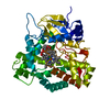
| ||||||||
|---|---|---|---|---|---|---|---|---|---|
| 1 |
| ||||||||
| Unit cell |
| ||||||||
| Atom site foot note | 1: CIS PROLINE - PRO 96 |
-Components
| #1: Protein | Mass: 44515.152 Da / Num. of mol.: 1 / Source method: isolated from a natural source / Source: (natural) Saccharopolyspora erythraea (bacteria) / References: UniProt: Q00441 |
|---|---|
| #2: Chemical | ChemComp-HEM /   Mass: 616.487 Da / Num. of mol.: 1 / Source method: obtained synthetically / Formula: C34H32FeN4O4 Mass: 616.487 Da / Num. of mol.: 1 / Source method: obtained synthetically / Formula: C34H32FeN4O4 |
| #3: Chemical | ChemComp-DEB / 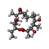  Mass: 386.523 Da / Num. of mol.: 1 / Source method: obtained synthetically / Formula: C21H38O6 Mass: 386.523 Da / Num. of mol.: 1 / Source method: obtained synthetically / Formula: C21H38O6 |
| #4: Water | ChemComp-HOH /  Mass: 18.015 Da / Num. of mol.: 241 / Source method: isolated from a natural source / Formula: H2O Mass: 18.015 Da / Num. of mol.: 241 / Source method: isolated from a natural source / Formula: H2O |
-Experimental details
-Experiment
| Experiment | Method: X-RAY DIFFRACTION / Number of used crystals: 1 |
|---|
- Sample preparation
| Crystal | Density Matthews: 2.41 Å3/Da / Density % sol: 48.95 % Description: TWO DERIVATIVE DATA SETS WERE ACQUIRED WITH A SIEMENS X-1000 AREA DETECTOR AND A ROTATING ANODE EQUIPPED WITH FOCUSING, MIRROR OPTICS. A NATIVE DATA SET WAS COLLECTED WITH TWO MARK III ...Description: TWO DERIVATIVE DATA SETS WERE ACQUIRED WITH A SIEMENS X-1000 AREA DETECTOR AND A ROTATING ANODE EQUIPPED WITH FOCUSING, MIRROR OPTICS. A NATIVE DATA SET WAS COLLECTED WITH TWO MARK III AREA DETECTORS AND A RIGAKU ROTATING ANODE AT THE UNIVERSITY OF CALIFORNIA, SAN DIEGO, U.S.A. | ||||||||||||||||||||
|---|---|---|---|---|---|---|---|---|---|---|---|---|---|---|---|---|---|---|---|---|---|
| Crystal grow | *PLUS pH: 8.5 / Method: vapor diffusion, hanging drop | ||||||||||||||||||||
| Components of the solutions | *PLUS
|
-Data collection
| Diffraction source | Wavelength: 1.5418 Å |
|---|---|
| Detector | Type: SIEMENS-NICOLET X100 / Detector: AREA DETECTOR |
| Radiation | Monochromatic (M) / Laue (L): M / Scattering type: x-ray |
| Radiation wavelength | Wavelength: 1.5418 Å / Relative weight: 1 |
| Reflection | Highest resolution: 2.1 Å / Num. obs: 23771 / % possible obs: 91 % / Observed criterion σ(I): 0 / Rmerge(I) obs: 0.0538 |
| Reflection | *PLUS Num. measured all: 127949 / Rmerge(I) obs: 0.0538 |
- Processing
| Software |
| ||||||||||||||||||||||||||||||||||||||||||||||||||||||||||||
|---|---|---|---|---|---|---|---|---|---|---|---|---|---|---|---|---|---|---|---|---|---|---|---|---|---|---|---|---|---|---|---|---|---|---|---|---|---|---|---|---|---|---|---|---|---|---|---|---|---|---|---|---|---|---|---|---|---|---|---|---|---|
| Refinement | Resolution: 2.1→10 Å / Rfactor Rwork: 0.196 / Rfactor obs: 0.196 / σ(F): 0 Details: RESIDUES 160 - 166 WERE NOT VISIBLE IN THE ELECTRON DENSITY. RESIDUE NUMBER 1 IS NOT VISIBLE. | ||||||||||||||||||||||||||||||||||||||||||||||||||||||||||||
| Refinement step | Cycle: LAST / Resolution: 2.1→10 Å
| ||||||||||||||||||||||||||||||||||||||||||||||||||||||||||||
| Refine LS restraints |
| ||||||||||||||||||||||||||||||||||||||||||||||||||||||||||||
| Software | *PLUS Name: X-PLOR / Classification: refinement | ||||||||||||||||||||||||||||||||||||||||||||||||||||||||||||
| Refinement | *PLUS | ||||||||||||||||||||||||||||||||||||||||||||||||||||||||||||
| Solvent computation | *PLUS | ||||||||||||||||||||||||||||||||||||||||||||||||||||||||||||
| Displacement parameters | *PLUS |
