 Movie
Movie Controller

[English] 日本語
 Yorodumi
- PDB-1ovh: T4 Lysozyme Cavity Mutant L99A/M102Q Bound With 2-Chloro-6-Methyl... -
+ Open data

- Basic information
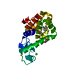
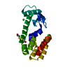
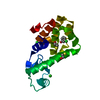
| Entry | Database: PDB / ID: 1ovh | ||||||
|---|---|---|---|---|---|---|---|
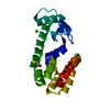
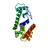
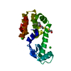
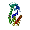
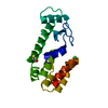
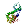
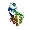
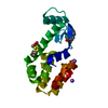
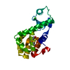
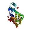
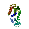
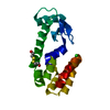
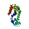
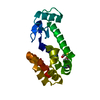
| Title | T4 Lysozyme Cavity Mutant L99A/M102Q Bound With 2-Chloro-6-Methyl-Aniline | ||||||
 Components Components | Lysozyme | ||||||
 Keywords Keywords | HYDROLASE / GLYCOSIDASE / BACTERIOLYTIC ENZYME | ||||||
| Function / homology |  Function and homology information Function and homology informationviral release from host cell by cytolysis / peptidoglycan catabolic process / cell wall macromolecule catabolic process / lysozyme / lysozyme activity / host cell cytoplasm / defense response to bacterium Similarity search - Function | ||||||
| Biological species |  Enterobacteria phage T4 (virus) Enterobacteria phage T4 (virus) | ||||||
| Method |  X-RAY DIFFRACTION / MOLECULAR REPLACEMENT / Resolution: 1.95 Å X-RAY DIFFRACTION / MOLECULAR REPLACEMENT / Resolution: 1.95 Å | ||||||
 Authors Authors | Wei, B.Q. / Baase, W.A. / Weaver, L.H. / Matthews, B.W. / Shoichet, B.K. | ||||||
 Citation Citation | Journal: J.Mol.Biol. / Year: 2004 Title: Testing a Flexible-receptor Docking Algorithm in a Model Binding Site Authors: Wei, B.Q. / Weaver, L.H. / Ferrari, A.M. / Matthews, B.W. / Shoichet, B.K. #1: Journal: J.Mol.Biol. / Year: 2002Title: A model Binding Site for Testing Scoring Functions in Molecular Docking Authors: Wei, B.Q. / Baase, W.A. / Weaver, L.H. / Matthews, B.W. / Shoichet, B.K. | ||||||
| History |
|
- Structure visualization
| Structure viewer | Molecule: MolmilJmol/JSmol |
|---|
- Downloads & links
-Download
| PDBx/mmCIF format | 1ovh.cif.gz | 47.6 KB | Display | PDBx/mmCIF format |
|---|---|---|---|---|
| PDB format | pdb1ovh.ent.gz | 32.6 KB | Display | PDB format |
| PDBx/mmJSON format | 1ovh.json.gz | Tree view | PDBx/mmJSON format | |
| Others |  Other downloads Other downloads |
-Validation report
| Arichive directory | https://data.pdbj.org/pub/pdb/validation_reports/ov/1ovhftp://data.pdbj.org/pub/pdb/validation_reports/ov/1ovh | HTTPS FTP |
|---|
-Related structure data
| Related structure data |  1ov5C 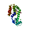 1ov7C 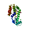 1ovjC 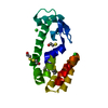 1ovkC 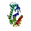 1owyC  1owzC 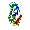 1lguS S: Starting model for refinement C: citing same article ( |
|---|---|
| Similar structure data |


-Links
 PDBj
PDBj


- Assembly
| Deposited unit | 
| ||||||||
|---|---|---|---|---|---|---|---|---|---|
| 1 |
| ||||||||
| Unit cell |
|
-Components
| #1: Protein | Mass: 18617.320 Da / Num. of mol.: 1 / Mutation: L99A, M102Q Source method: isolated from a genetically manipulated source Source: (gene. exp.) Enterobacteria phage T4 (virus) / Genus: T4-like viruses / Species: Enterobacteria phage T4 sensu lato / Production host:  | ||||||
|---|---|---|---|---|---|---|---|
| #2: Chemical |   Mass: 35.453 Da / Num. of mol.: 2 / Source method: obtained synthetically / Formula: Cl Mass: 35.453 Da / Num. of mol.: 2 / Source method: obtained synthetically / Formula: Cl#3: Chemical |   Mass: 78.133 Da / Num. of mol.: 3 / Source method: obtained synthetically / Formula: C2H6OS Mass: 78.133 Da / Num. of mol.: 3 / Source method: obtained synthetically / Formula: C2H6OS#4: Chemical | ChemComp-2CM / |   Mass: 141.598 Da / Num. of mol.: 1 / Source method: obtained synthetically / Formula: C7H8ClN Mass: 141.598 Da / Num. of mol.: 1 / Source method: obtained synthetically / Formula: C7H8ClN#5: Water | ChemComp-HOH / |  Mass: 18.015 Da / Num. of mol.: 62 / Source method: isolated from a natural source / Formula: H2O Mass: 18.015 Da / Num. of mol.: 62 / Source method: isolated from a natural source / Formula: H2O |
-Experimental details
-Experiment
| Experiment | Method: X-RAY DIFFRACTION / Number of used crystals: 1 |
|---|
- Sample preparation
| Crystal | Density Matthews: 2.68 Å3/Da / Density % sol: 53.8 % |
|---|
-Data collection
| Diffraction | Mean temperature: 298 K |
|---|---|
| Diffraction source | Source: ROTATING ANODE / Type: RIGAKU RU200 / Wavelength: 1.5418 Å |
| Detector | Type: UCSD MARK II / Detector: AREA DETECTOR |
| Radiation | Monochromator: graphite / Protocol: SINGLE WAVELENGTH / Monochromatic (M) / Laue (L): M / Scattering type: x-ray |
| Radiation wavelength | Wavelength: 1.5418 Å / Relative weight: 1 |
| Reflection | Resolution: 1.95→13 Å / Num. all: 14679 / Num. obs: 14679 / % possible obs: 93 % / Observed criterion σ(F): 0 / Observed criterion σ(I): 0 / Rmerge(I) obs: 0.08 / Net I/σ(I): 11.2 |
| Reflection shell | Resolution: 1.95→2.1 Å / Rmerge(I) obs: 0.161 / Mean I/σ(I) obs: 2.3 / Num. unique all: 2676 / % possible all: 87 |
- Processing
| Software |
| |||||||||||||||||||||
|---|---|---|---|---|---|---|---|---|---|---|---|---|---|---|---|---|---|---|---|---|---|---|
| Refinement | Method to determine structure: MOLECULAR REPLACEMENT Starting model: PDB entry 1LGU Resolution: 1.95→13 Å / Isotropic thermal model: isotropic / σ(F): 0 / σ(I): 0 / Stereochemistry target values: Engh & Huber
| |||||||||||||||||||||
| Refinement step | Cycle: LAST / Resolution: 1.95→13 Å
| |||||||||||||||||||||
| Refine LS restraints |
|
