 Movie
Movie Controller


+ Open data

- Basic information
| Entry | Database: PDB / ID: 1otk | ||||||
|---|---|---|---|---|---|---|---|
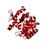
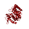
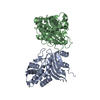
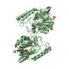
| Title | Structural Genomics, Protein paaC | ||||||
 Components Components | Phenylacetic acid degradation protein paaC | ||||||
 Keywords Keywords | OXIDOREDUCTASE / STRUCTURAL GENOMICS / PSI / Protein Structure Initiative / Midwest Center for Structural Genomics / MCSG | ||||||
| Function / homology |  Function and homology information Function and homology informationphenylacetyl-CoA 1,2-epoxidase complex / phenylacetate catabolic process / cytosol Similarity search - Function | ||||||
| Biological species |  | ||||||
| Method |  X-RAY DIFFRACTION / SYNCHROTRON / SAD / Resolution: 2 Å X-RAY DIFFRACTION / SYNCHROTRON / SAD / Resolution: 2 Å | ||||||
 Authors Authors | Zhang, R. / Joachimiak, A. / Edwards, A. / Savchenko, A. / Skarina, T. / Midwest Center for Structural Genomics (MCSG) | ||||||
 Citation Citation | Journal: To be Published Title: The 2 A crystal structure of protein paaC from E. Coli Authors: Zhang, R. / Joachimiak, A. / Edwards, A. / Savchenko, A. / Skarina, T. | ||||||
| History |
|
- Structure visualization
| Structure viewer | Molecule: MolmilJmol/JSmol |
|---|
- Downloads & links
-Download
| PDBx/mmCIF format | 1otk.cif.gz | 111.4 KB | Display | PDBx/mmCIF format |
|---|---|---|---|---|
| PDB format | pdb1otk.ent.gz | 86.4 KB | Display | PDB format |
| PDBx/mmJSON format | 1otk.json.gz | Tree view | PDBx/mmJSON format | |
| Others |  Other downloads Other downloads |
-Validation report
| Arichive directory | https://data.pdbj.org/pub/pdb/validation_reports/ot/1otkftp://data.pdbj.org/pub/pdb/validation_reports/ot/1otk | HTTPS FTP |
|---|
-Related structure data
| Similar structure data | |
|---|---|
| Other databases |


-Links
 PDBj
PDBj


- Assembly
| Deposited unit | 
| ||||||||
|---|---|---|---|---|---|---|---|---|---|
| 1 |
| ||||||||
| Unit cell |
| ||||||||
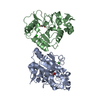
| Details | This protein exists as dimer which consists of molA and molB. |
-Components
| #1: Protein | Mass: 27969.404 Da / Num. of mol.: 2 Source method: isolated from a genetically manipulated source Source: (gene. exp.) #2: Water | ChemComp-HOH / |  Mass: 18.015 Da / Num. of mol.: 342 / Source method: isolated from a natural source / Formula: H2O Mass: 18.015 Da / Num. of mol.: 342 / Source method: isolated from a natural source / Formula: H2OHas protein modification | Y | |
|---|
-Experimental details
-Experiment
| Experiment | Method: X-RAY DIFFRACTION / Number of used crystals: 1 |
|---|
- Sample preparation
| Crystal | Density Matthews: 3.22 Å3/Da / Density % sol: 61.78 % |
|---|---|
| Crystal grow | Temperature: 298 K / Method: vapor diffusion, hanging drop / pH: 6.5 Details: 11% iso-Prop., 0.1 M Nacacod., 0.2M NaCitr., pH 6.5, VAPOR DIFFUSION, HANGING DROP, temperature 298K |
-Data collection
| Diffraction | Mean temperature: 100 K |
|---|---|
| Diffraction source | Source: SYNCHROTRON / Site: APS  / Beamline: 19-ID / Wavelength: 0.9793 Å / Beamline: 19-ID / Wavelength: 0.9793 Å |
| Detector | Type: SBC-2 / Detector: CCD / Date: Mar 17, 2003 / Details: Si 111 Channel |
| Radiation | Monochromator: Si 111 Channel / Protocol: SINGLE WAVELENGTH / Monochromatic (M) / Laue (L): M / Scattering type: x-ray |
| Radiation wavelength | Wavelength: 0.9793 Å / Relative weight: 1 |
| Reflection | Resolution: 2→50 Å / Num. all: 49792 / Num. obs: 49742 / % possible obs: 99.9 % / Observed criterion σ(F): 2 / Observed criterion σ(I): 2 / Redundancy: 10.76 % / Biso Wilson estimate: 12.7 Å2 / Rmerge(I) obs: 0.113 / Net I/σ(I): 33.1 |
| Reflection shell | Resolution: 2→2.07 Å / Redundancy: 5.6 % / Rmerge(I) obs: 0.52 / Mean I/σ(I) obs: 5.84 / Num. unique all: 4898 / % possible all: 98.9 |
- Processing
| Software |
| ||||||||||||||||||||
|---|---|---|---|---|---|---|---|---|---|---|---|---|---|---|---|---|---|---|---|---|---|
| Refinement | Method to determine structure: SAD / Resolution: 2→46.82 Å / Rfactor Rfree error: 0.003 / Isotropic thermal model: RESTRAINED / Cross valid method: THROUGHOUT / σ(F): 0 Details: The number of reflections for refinement includes Freidel pairs.
| ||||||||||||||||||||
| Solvent computation | Solvent model: FLAT MODEL / Bsol: 50.7366 Å2 / ksol: 0.34617 e/Å3 | ||||||||||||||||||||
| Displacement parameters | Biso mean: 22.9 Å2
| ||||||||||||||||||||
| Refine analyze | Luzzati coordinate error free: 0.25 Å / Luzzati sigma a free: 0.18 Å | ||||||||||||||||||||
| Refinement step | Cycle: LAST / Resolution: 2→46.82 Å
| ||||||||||||||||||||
| Refine LS restraints |
| ||||||||||||||||||||
| LS refinement shell | Resolution: 2→2.13 Å / Rfactor Rfree error: 0.009 / Total num. of bins used: 6
| ||||||||||||||||||||
| Xplor file |
|
