 Movie
Movie Controller

[English] 日本語
 Yorodumi
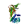
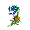
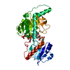
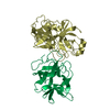
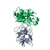
- PDB-1ori: Structure of the predominant protein arginine methyltransferase PRMT1 -
+ Open data

- Basic information
| Entry | Database: PDB / ID: 1ori | ||||||
|---|---|---|---|---|---|---|---|
| Title | Structure of the predominant protein arginine methyltransferase PRMT1 | ||||||
 Components Components | Protein arginine N-methyltransferase 1 | ||||||
 Keywords Keywords | TRANSFERASE / protein arginine methylation AdoMet-dependent methylation | ||||||
| Function / homology |  Function and homology information Function and homology informationsnoRNP binding / peptidyl-arginine omega-N-methylation / RUNX1 regulates genes involved in megakaryocyte differentiation and platelet function / peptidyl-arginine methylation, to asymmetrical-dimethyl arginine / Estrogen-dependent gene expression / RMTs methylate histone arginines / GATOR1 complex binding / Extra-nuclear estrogen signaling / positive regulation of hemoglobin biosynthetic process / protein-arginine omega-N monomethyltransferase activity ...snoRNP binding / peptidyl-arginine omega-N-methylation / RUNX1 regulates genes involved in megakaryocyte differentiation and platelet function / peptidyl-arginine methylation, to asymmetrical-dimethyl arginine / Estrogen-dependent gene expression / RMTs methylate histone arginines / GATOR1 complex binding / Extra-nuclear estrogen signaling / positive regulation of hemoglobin biosynthetic process / protein-arginine omega-N monomethyltransferase activity / N-methyltransferase activity / regulation of BMP signaling pathway / protein-arginine omega-N asymmetric methyltransferase activity / S-adenosylmethionine metabolic process / regulation of megakaryocyte differentiation / type I protein arginine methyltransferase / histone H4R3 methyltransferase activity / protein methyltransferase activity / protein methylation / S-adenosylmethionine-dependent methyltransferase activity / protein-arginine N-methyltransferase activity / cellular response to methionine / methylosome / S-adenosyl-L-methionine binding / methyl-CpG binding / positive regulation of p38MAPK cascade / cardiac muscle tissue development / negative regulation of JNK cascade / histone methyltransferase activity / mitogen-activated protein kinase p38 binding / negative regulation of megakaryocyte differentiation / positive regulation of double-strand break repair via homologous recombination / positive regulation of TORC1 signaling / liver regeneration / RNA splicing / positive regulation of erythrocyte differentiation / positive regulation of translation / protein homooligomerization / neuron projection development / in utero embryonic development / chromatin remodeling / lysosomal membrane / positive regulation of cell population proliferation / DNA damage response / regulation of DNA-templated transcription / enzyme binding / protein-containing complex / nucleoplasm / identical protein binding / nucleus / cytosol / cytoplasm Similarity search - Function | ||||||
| Biological species |  | ||||||
| Method |  X-RAY DIFFRACTION / SYNCHROTRON / MOLECULAR REPLACEMENT / Resolution: 2.5 Å X-RAY DIFFRACTION / SYNCHROTRON / MOLECULAR REPLACEMENT / Resolution: 2.5 Å | ||||||
 Authors Authors | Zhang, X. / Cheng, X. | ||||||
 Citation Citation | Journal: Structure / Year: 2003 Title: Structure of the Predominant Protein Arginine Methyltransferase PRMT1 and Analysis of its Binding to Substrate Peptides Authors: Zhang, X. / Cheng, X. | ||||||
| History |
|
- Structure visualization
| Structure viewer | Molecule: MolmilJmol/JSmol |
|---|
- Downloads & links
-Download
| PDBx/mmCIF format | 1ori.cif.gz | 81.9 KB | Display | PDBx/mmCIF format |
|---|---|---|---|---|
| PDB format | pdb1ori.ent.gz | 59.3 KB | Display | PDB format |
| PDBx/mmJSON format | 1ori.json.gz | Tree view | PDBx/mmJSON format | |
| Others |  Other downloads Other downloads |
-Validation report
| Summary document | 1ori_validation.pdf.gz | 449.2 KB | Display | wwPDB validaton report |
|---|---|---|---|---|
| Full document | 1ori_full_validation.pdf.gz | 457.5 KB | Display | |
| Data in XML | 1ori_validation.xml.gz | 8.9 KB | Display | |
| Data in CIF | 1ori_validation.cif.gz | 13.3 KB | Display | |
| Arichive directory | https://data.pdbj.org/pub/pdb/validation_reports/or/1oriftp://data.pdbj.org/pub/pdb/validation_reports/or/1ori | HTTPS FTP |
-Related structure data
| Related structure data | 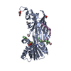 1or8C 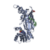 1orhC 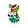 1f3lS C: citing same article ( S: Starting model for refinement |
|---|---|
| Similar structure data |


-Links
 PDBj
PDBj


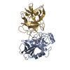
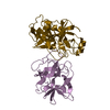
- Assembly
| Deposited unit | 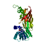
| ||||||||
|---|---|---|---|---|---|---|---|---|---|
| 1 |
| ||||||||
| 2 | 
| ||||||||
| Unit cell |
| ||||||||
| Components on special symmetry positions |
|
-Components
| #1: Protein | Mass: 39627.297 Da / Num. of mol.: 1 / Fragment: M11 Source method: isolated from a genetically manipulated source Source: (gene. exp.)  | ||
|---|---|---|---|
| #2: Chemical | ChemComp-SAH /   Type: L-peptide linking / Mass: 384.411 Da / Num. of mol.: 1 / Source method: obtained synthetically / Formula: C14H20N6O5S Type: L-peptide linking / Mass: 384.411 Da / Num. of mol.: 1 / Source method: obtained synthetically / Formula: C14H20N6O5S | ||
| #3: Chemical | Num. of mol.: 2 / Source method: obtained synthetically #4: Water | ChemComp-HOH / |  Mass: 18.015 Da / Num. of mol.: 129 / Source method: isolated from a natural source / Formula: H2O Mass: 18.015 Da / Num. of mol.: 129 / Source method: isolated from a natural source / Formula: H2O |
-Experimental details
-Experiment
| Experiment | Method: X-RAY DIFFRACTION / Number of used crystals: 1 |
|---|
- Sample preparation
| Crystal | Density Matthews: 3.57 Å3/Da / Density % sol: 65.51 % | ||||||||||||||||||||||||
|---|---|---|---|---|---|---|---|---|---|---|---|---|---|---|---|---|---|---|---|---|---|---|---|---|---|
| Crystal grow | Temperature: 289 K / Method: vapor diffusion, hanging drop / pH: 4.7 Details: ammonium phosphate, pH 4.7, VAPOR DIFFUSION, HANGING DROP, temperature 289K | ||||||||||||||||||||||||
| Crystal grow | *PLUS Method: vapor diffusion, hanging drop | ||||||||||||||||||||||||
| Components of the solutions | *PLUS
|
-Data collection
| Diffraction | Mean temperature: 100 K |
|---|---|
| Diffraction source | Source: SYNCHROTRON / Site: NSLS  / Beamline: X26C / Wavelength: 1.1 Å / Beamline: X26C / Wavelength: 1.1 Å |
| Detector | Type: ADSC QUANTUM 4 / Detector: CCD / Date: Mar 31, 2000 |
| Radiation | Protocol: SINGLE WAVELENGTH / Monochromatic (M) / Laue (L): M / Scattering type: x-ray |
| Radiation wavelength | Wavelength: 1.1 Å / Relative weight: 1 |
| Reflection | Resolution: 2.5→25 Å / Num. all: 20200 / Num. obs: 20200 / % possible obs: 98.6 % / Observed criterion σ(F): 0 / Observed criterion σ(I): -3 / Redundancy: 5.07 % / Biso Wilson estimate: 34.5 Å2 / Rmerge(I) obs: 0.1 / Net I/σ(I): 19.2 |
| Reflection shell | Resolution: 2.5→2.54 Å / Rmerge(I) obs: 0.213 / Mean I/σ(I) obs: 6.7 / Num. unique all: 952 / % possible all: 96.2 |
| Reflection | *PLUS Highest resolution: 2.5 Å / Num. measured all: 102483 / Rmerge(I) obs: 0.1 |
- Processing
| Software |
| |||||||||||||||||||||||||
|---|---|---|---|---|---|---|---|---|---|---|---|---|---|---|---|---|---|---|---|---|---|---|---|---|---|---|
| Refinement | Method to determine structure: MOLECULAR REPLACEMENT Starting model: PDB entry 1F3L Resolution: 2.5→24.18 Å / Isotropic thermal model: RESTRAINED / Cross valid method: THROUGHOUT / σ(F): 2 / Details: BULK SOLVENT MODEL USED
| |||||||||||||||||||||||||
| Displacement parameters | Biso mean: 38.7 Å2
| |||||||||||||||||||||||||
| Refine analyze |
| |||||||||||||||||||||||||
| Refinement step | Cycle: LAST / Resolution: 2.5→24.18 Å
| |||||||||||||||||||||||||
| Refine LS restraints |
| |||||||||||||||||||||||||
| LS refinement shell | Resolution: 2.5→2.61 Å / Rfactor Rfree error: 0.028
| |||||||||||||||||||||||||
| Refinement | *PLUS | |||||||||||||||||||||||||
| Solvent computation | *PLUS | |||||||||||||||||||||||||
| Displacement parameters | *PLUS | |||||||||||||||||||||||||
| Refine LS restraints | *PLUS
|
