 Movie
Movie Controller

[English] 日本語
 Yorodumi
- PDB-1ooe: Structural Genomics of Caenorhabditis elegans : Dihydropteridine ... -
+ Open data

- Basic information
| Entry | Database: PDB / ID: 1ooe | ||||||
|---|---|---|---|---|---|---|---|
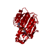
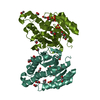
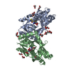
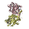
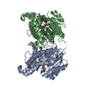
| Title | Structural Genomics of Caenorhabditis elegans : Dihydropteridine reductase | ||||||
 Components Components | Dihydropteridine reductase | ||||||
 Keywords Keywords | OXIDOREDUCTASE / Structural Genomics / Dihydropteridine reductase / PSI / Protein Structure Initiative / Southeast Collaboratory for Structural Genomics / SECSG | ||||||
| Function / homology |  Function and homology information Function and homology informationPhenylalanine metabolism / 6,7-dihydropteridine reductase / 6,7-dihydropteridine reductase activity / NADH binding / tetrahydrobiopterin biosynthetic process / L-phenylalanine catabolic process / NADPH binding / cytoplasm Similarity search - Function | ||||||
| Biological species |  | ||||||
| Method |  X-RAY DIFFRACTION / SYNCHROTRON / MOLECULAR REPLACEMENT / Resolution: 1.65 Å X-RAY DIFFRACTION / SYNCHROTRON / MOLECULAR REPLACEMENT / Resolution: 1.65 Å | ||||||
 Authors Authors | Symersky, J. / Li, S. / Nagy, L. / Qiu, S. / Lin, G. / Tsao, J. / Luo, D. / Carson, M. / DeLucas, L. / Luo, M. / Southeast Collaboratory for Structural Genomics (SECSG) | ||||||
 Citation Citation | Journal: Proteins / Year: 2003 Title: Structural genomics of Caenorhabditis elegans: structure of dihydropteridine reductase. Authors: Symersky, J. / Li, S. / Carson, M. / Luo, D. / Luan, C.H. / Luo, M. | ||||||
| History |
|
- Structure visualization
| Structure viewer | Molecule: MolmilJmol/JSmol |
|---|
- Downloads & links
-Download
| PDBx/mmCIF format | 1ooe.cif.gz | 110.6 KB | Display | PDBx/mmCIF format |
|---|---|---|---|---|
| PDB format | pdb1ooe.ent.gz | 85 KB | Display | PDB format |
| PDBx/mmJSON format | 1ooe.json.gz | Tree view | PDBx/mmJSON format | |
| Others |  Other downloads Other downloads |
-Validation report
| Arichive directory | https://data.pdbj.org/pub/pdb/validation_reports/oo/1ooeftp://data.pdbj.org/pub/pdb/validation_reports/oo/1ooe | HTTPS FTP |
|---|
-Related structure data
| Related structure data |  1dhrS S: Starting model for refinement |
|---|---|
| Similar structure data | |
| Other databases |


-Links
 PDBj
PDBj


- Assembly
| Deposited unit | 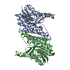
| ||||||||
|---|---|---|---|---|---|---|---|---|---|
| 1 |
| ||||||||
| Unit cell |
|
-Components
| #1: Protein | Mass: 24734.938 Da / Num. of mol.: 2 Source method: isolated from a genetically manipulated source Source: (gene. exp.)  #2: Chemical |   Mass: 195.237 Da / Num. of mol.: 2 / Source method: obtained synthetically / Formula: C6H13NO4S / Comment: pH buffer*YM Mass: 195.237 Da / Num. of mol.: 2 / Source method: obtained synthetically / Formula: C6H13NO4S / Comment: pH buffer*YM#3: Water | ChemComp-HOH / |  Mass: 18.015 Da / Num. of mol.: 601 / Source method: isolated from a natural source / Formula: H2O Mass: 18.015 Da / Num. of mol.: 601 / Source method: isolated from a natural source / Formula: H2O |
|---|
-Experimental details
-Experiment
| Experiment | Method: X-RAY DIFFRACTION / Number of used crystals: 1 |
|---|
- Sample preparation
| Crystal | Density Matthews: 2.05 Å3/Da / Density % sol: 39.5 % | |||||||||||||||||||||||||||||||||||||||||||||||||
|---|---|---|---|---|---|---|---|---|---|---|---|---|---|---|---|---|---|---|---|---|---|---|---|---|---|---|---|---|---|---|---|---|---|---|---|---|---|---|---|---|---|---|---|---|---|---|---|---|---|---|
| Crystal grow | Temperature: 295 K / Method: vapor diffusion, hanging drop / pH: 5.6 Details: pH 5.6, VAPOR DIFFUSION, HANGING DROP, temperature 295.0K RESERVOIR: 20% PEG8000, 10 MM MGCL2, 0.1 M AMMONIUM SULFATE, 50 MM MES, PH 5.6; PROTEIN STOCK: 17.3 MG/ML IN 10 MM HEPES, PH 7.0; ...Details: pH 5.6, VAPOR DIFFUSION, HANGING DROP, temperature 295.0K RESERVOIR: 20% PEG8000, 10 MM MGCL2, 0.1 M AMMONIUM SULFATE, 50 MM MES, PH 5.6; PROTEIN STOCK: 17.3 MG/ML IN 10 MM HEPES, PH 7.0; DROPS: 2 MICROLITERS OF PROTEIN STOCK + 4 MICROLITERS OF RESERVOIR SOLUTION | |||||||||||||||||||||||||||||||||||||||||||||||||
| Crystal grow | *PLUS Temperature: 295 K / pH: 7 | |||||||||||||||||||||||||||||||||||||||||||||||||
| Components of the solutions | *PLUS
|
-Data collection
| Diffraction | Mean temperature: 100 K |
|---|---|
| Diffraction source | Source: SYNCHROTRON / Site: APS  / Beamline: 22-ID / Wavelength: 1.07175 Å / Beamline: 22-ID / Wavelength: 1.07175 Å |
| Detector | Type: MAR CCD 165 mm / Detector: CCD / Date: Nov 3, 2002 |
| Radiation | Protocol: SINGLE WAVELENGTH / Monochromatic (M) / Laue (L): M / Scattering type: x-ray |
| Radiation wavelength | Wavelength: 1.07175 Å / Relative weight: 1 |
| Reflection | Resolution: 1.6→50 Å / Num. all: 56662 / Num. obs: 56662 / % possible obs: 93.9 % / Observed criterion σ(F): 0 / Observed criterion σ(I): -3 / Redundancy: 1.9 % / Biso Wilson estimate: 18.1 Å2 / Rsym value: 0.041 / Net I/σ(I): 10 |
| Reflection shell | Resolution: 1.6→1.64 Å / Redundancy: 0.8 % / Mean I/σ(I) obs: 3.1 / Num. unique all: 2969 / Rsym value: 0.281 / % possible all: 74 |
| Reflection | *PLUS Highest resolution: 1.65 Å / Lowest resolution: 32.28 Å / % possible obs: 95.3 % / Num. measured all: 102854 / Rmerge(I) obs: 0.041 |
| Reflection shell | *PLUS Highest resolution: 1.65 Å / Lowest resolution: 1.71 Å / % possible obs: 93.8 % / Rmerge(I) obs: 0.281 |
- Processing
| Software |
| ||||||||||||||||||||||||||||||||||||
|---|---|---|---|---|---|---|---|---|---|---|---|---|---|---|---|---|---|---|---|---|---|---|---|---|---|---|---|---|---|---|---|---|---|---|---|---|---|
| Refinement | Method to determine structure: MOLECULAR REPLACEMENT Starting model: PDB entry 1DHR Resolution: 1.65→32.28 Å / Rfactor Rfree error: 0.004 / Data cutoff high absF: 10000 / Data cutoff high rms absF: 10000 / Isotropic thermal model: RESTRAINED / Cross valid method: THROUGHOUT / σ(F): 0 / Stereochemistry target values: Engh & Huber Details: THE ELECTRON DENSITY AND RSR VALUE SUPPORT THE CONFORMATION OUTSIDE THE EXPECTED RAMACHANDRAN REGIONS.
| ||||||||||||||||||||||||||||||||||||
| Solvent computation | Solvent model: FLAT MODEL / Bsol: 36.9819 Å2 / ksol: 0.294278 e/Å3 | ||||||||||||||||||||||||||||||||||||
| Displacement parameters | Biso mean: 16.1 Å2 | ||||||||||||||||||||||||||||||||||||
| Refine analyze |
| ||||||||||||||||||||||||||||||||||||
| Refinement step | Cycle: LAST / Resolution: 1.65→32.28 Å
| ||||||||||||||||||||||||||||||||||||
| Refine LS restraints |
| ||||||||||||||||||||||||||||||||||||
| LS refinement shell | Resolution: 1.65→1.71 Å / Rfactor Rfree error: 0.017 / Total num. of bins used: 10
| ||||||||||||||||||||||||||||||||||||
| Xplor file |
| ||||||||||||||||||||||||||||||||||||
| Refinement | *PLUS % reflection Rfree: 5 % | ||||||||||||||||||||||||||||||||||||
| Solvent computation | *PLUS | ||||||||||||||||||||||||||||||||||||
| Displacement parameters | *PLUS | ||||||||||||||||||||||||||||||||||||
| Refine LS restraints | *PLUS
|
