 Movie
Movie Controller

[English] 日本語
 Yorodumi
- PDB-1ofw: Three dimensional structure of the oxidized form of nine heme cyt... -
+ Open data

- Basic information
| Entry | Database: PDB / ID: 1ofw | ||||||
|---|---|---|---|---|---|---|---|
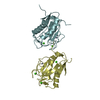
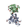
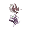
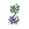
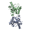
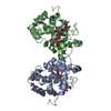
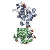
| Title | Three dimensional structure of the oxidized form of nine heme cytochrome c at PH 7.5 | ||||||
 Components Components | NINE-HEME CYTOCHROME C | ||||||
 Keywords Keywords | ELECTRON TRANSPORT / MULTIHEME CYTOCHROME C / ELECTRON TRANSFER / ELECTRON TRANSPOR | ||||||
| Function / homology |  Function and homology information Function and homology informationperiplasmic space / electron transfer activity / heme binding / metal ion binding Similarity search - Function | ||||||
| Biological species |  DESULFOVIBRIO DESULFURICANS (bacteria) DESULFOVIBRIO DESULFURICANS (bacteria) | ||||||
| Method |  X-RAY DIFFRACTION / SYNCHROTRON / MOLECULAR REPLACEMENT / Resolution: 1.5 Å X-RAY DIFFRACTION / SYNCHROTRON / MOLECULAR REPLACEMENT / Resolution: 1.5 Å | ||||||
 Authors Authors | Bento, I. / Teixeira, V.H. / Baptista, A.M. / Soares, C.M. / Matias, P.M. / Carrondo, M.A. | ||||||
 Citation Citation | Journal: J.Biol.Chem. / Year: 2003 Title: Redox-Bohr and Other Cooperativity Effects in the Nine-Heme Cytochrome C from Desulfovibrio Desulfuricans Atcc 27774: Crystallographic and Modeling Studies Authors: Bento, I. / Teixeira, V.H. / Baptista, A.M. / Soares, C.M. / Matias, P.M. / Carrondo, M.A. | ||||||
| History |
|
- Structure visualization
| Structure viewer | Molecule: MolmilJmol/JSmol |
|---|
- Downloads & links
-Download
| PDBx/mmCIF format | 1ofw.cif.gz | 164.6 KB | Display | PDBx/mmCIF format |
|---|---|---|---|---|
| PDB format | pdb1ofw.ent.gz | 131.7 KB | Display | PDB format |
| PDBx/mmJSON format | 1ofw.json.gz | Tree view | PDBx/mmJSON format | |
| Others |  Other downloads Other downloads |
-Validation report
| Arichive directory | https://data.pdbj.org/pub/pdb/validation_reports/of/1ofwftp://data.pdbj.org/pub/pdb/validation_reports/of/1ofw | HTTPS FTP |
|---|
-Related structure data
| Related structure data |  1ofyC 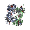 19hcS S: Starting model for refinement C: citing same article ( |
|---|---|
| Similar structure data |


-Links
 PDBj
PDBj


- Assembly
| Deposited unit | 
| ||||||||
|---|---|---|---|---|---|---|---|---|---|
| 1 | 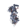
| ||||||||
| 2 | 
| ||||||||
| Unit cell |
|
-Components
| #1: Protein | Mass: 32264.016 Da / Num. of mol.: 2 / Source method: isolated from a natural source / Source: (natural) DESULFOVIBRIO DESULFURICANS (bacteria) / References: UniProt: Q9RN68#2: Chemical | ChemComp-HEC /   Mass: 618.503 Da / Num. of mol.: 18 / Source method: obtained synthetically / Formula: C34H34FeN4O4 Mass: 618.503 Da / Num. of mol.: 18 / Source method: obtained synthetically / Formula: C34H34FeN4O4#3: Chemical | ChemComp-GOL /   Mass: 92.094 Da / Num. of mol.: 4 / Source method: obtained synthetically / Formula: C3H8O3 Mass: 92.094 Da / Num. of mol.: 4 / Source method: obtained synthetically / Formula: C3H8O3#4: Chemical | ChemComp-ACT / |   Mass: 59.044 Da / Num. of mol.: 1 / Source method: obtained synthetically / Formula: C2H3O2 Mass: 59.044 Da / Num. of mol.: 1 / Source method: obtained synthetically / Formula: C2H3O2#5: Water | ChemComp-HOH / |  Mass: 18.015 Da / Num. of mol.: 641 / Source method: isolated from a natural source / Formula: H2O Mass: 18.015 Da / Num. of mol.: 641 / Source method: isolated from a natural source / Formula: H2OCompound details | COULD FORM PART OF A TRANSMEMBRANE REDOX COMPLEX THROUGH WHICH ELECTRONS ARE TRANSFERRED TO THE ...COULD FORM PART OF A TRANSMEMBR | Has protein modification | Y | |
|---|
-Experimental details
-Experiment
| Experiment | Method: X-RAY DIFFRACTION / Number of used crystals: 1 |
|---|
- Sample preparation
| Crystal | Density Matthews: 3.29 Å3/Da / Density % sol: 62.6 % | ||||||||||||||||||||||||
|---|---|---|---|---|---|---|---|---|---|---|---|---|---|---|---|---|---|---|---|---|---|---|---|---|---|
| Crystal grow | pH: 7.5 / Details: pH 7.50 | ||||||||||||||||||||||||
| Crystal grow | *PLUS pH: 7.5 / Method: vapor diffusion, sitting drop / Details: Coelho, A.V., (1996) Protein Sci., 5, 1189. | ||||||||||||||||||||||||
| Components of the solutions | *PLUS
|
-Data collection
| Diffraction | Mean temperature: 110 K |
|---|---|
| Diffraction source | Source: SYNCHROTRON / Site: ESRF  / Beamline: ID14-2 / Wavelength: 0.933 / Beamline: ID14-2 / Wavelength: 0.933 |
| Detector | Type: ADSC CCD / Detector: CCD / Details: TOROIDAL MIRROR |
| Radiation | Monochromator: DIAMOND (111) CRYSTAL / Protocol: SINGLE WAVELENGTH / Monochromatic (M) / Laue (L): M / Scattering type: x-ray |
| Radiation wavelength | Wavelength: 0.933 Å / Relative weight: 1 |
| Reflection | Resolution: 1.5→25 Å / Num. obs: 153684 / % possible obs: 98.2 % / Redundancy: 2.14 % / Rmerge(I) obs: 0.036 / Net I/σ(I): 19 |
| Reflection shell | Resolution: 1.5→1.55 Å / Redundancy: 2.14 % / Rmerge(I) obs: 0.269 / Mean I/σ(I) obs: 3 / % possible all: 96.8 |
| Reflection | *PLUS Highest resolution: 1.5 Å / Lowest resolution: 25 Å / Redundancy: 2.14 % / Num. measured all: 330192 / Rmerge(I) obs: 0.036 |
| Reflection shell | *PLUS % possible obs: 96.8 % / Rmerge(I) obs: 0.269 |
- Processing
| Software |
| |||||||||||||||||||||||||||||||||
|---|---|---|---|---|---|---|---|---|---|---|---|---|---|---|---|---|---|---|---|---|---|---|---|---|---|---|---|---|---|---|---|---|---|---|
| Refinement | Method to determine structure: MOLECULAR REPLACEMENT Starting model: 19HC Resolution: 1.5→25 Å / Num. parameters: 24267 / Num. restraintsaints: 29987 / Cross valid method: THROUGHOUT / σ(F): 0 / Stereochemistry target values: ENGH AND HUBER
| |||||||||||||||||||||||||||||||||
| Solvent computation | Solvent model: MOEWS & KRETSINGER | |||||||||||||||||||||||||||||||||
| Refine analyze | Num. disordered residues: 23 / Occupancy sum non hydrogen: 5837.12 | |||||||||||||||||||||||||||||||||
| Refinement step | Cycle: LAST / Resolution: 1.5→25 Å
| |||||||||||||||||||||||||||||||||
| Refine LS restraints |
| |||||||||||||||||||||||||||||||||
| Software | *PLUS Name: SHELXL / Version: 97 / Classification: refinement | |||||||||||||||||||||||||||||||||
| Refinement | *PLUS Rfactor Rfree: 0.193 / Rfactor Rwork: 0.161 | |||||||||||||||||||||||||||||||||
| Solvent computation | *PLUS | |||||||||||||||||||||||||||||||||
| Displacement parameters | *PLUS |
