 Movie
Movie Controller


+ Open data

- Basic information
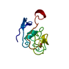
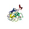
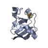
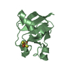
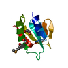
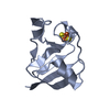
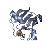
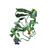
| Entry | Database: PDB / ID: 1off | ||||||
|---|---|---|---|---|---|---|---|
| Title | 2Fe-2S Ferredoxin from Synechocystis sp. PCC 6803 | ||||||
 Components Components | FERREDOXIN I | ||||||
 Keywords Keywords | ELECTRON TRANSPORT / FERREDOXIN / ELECTRON TRANSFER / IRON-SULPHUR / 2FE-2S / METAL-BINDING | ||||||
| Function / homology |  Function and homology information Function and homology informationelectron transport chain / 2 iron, 2 sulfur cluster binding / electron transfer activity / metal ion binding Similarity search - Function | ||||||
| Biological species |  | ||||||
| Method |  X-RAY DIFFRACTION / MOLECULAR REPLACEMENT / Resolution: 1.8 Å X-RAY DIFFRACTION / MOLECULAR REPLACEMENT / Resolution: 1.8 Å | ||||||
 Authors Authors | Van Den heuvel, R.H.H. / Mattevi, A. | ||||||
 Citation Citation | Journal: J.Mol.Biol. / Year: 2003 Title: The Active Conformation of Glutamate Synthase and its Binding to Ferredoxin Authors: Van Den Heuvel, R.H.H. / Svergun, D.I. / Petoukhov, M.V. / Coda, A. / Curti, B. / Ravasio, S. / Vanoni, M.A. / Mattevi, A. | ||||||
| History |
|
- Structure visualization
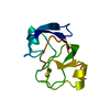
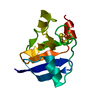
| Structure viewer | Molecule: MolmilJmol/JSmol |
|---|
- Downloads & links
-Download
| PDBx/mmCIF format | 1off.cif.gz | 32.8 KB | Display | PDBx/mmCIF format |
|---|---|---|---|---|
| PDB format | pdb1off.ent.gz | 21.5 KB | Display | PDB format |
| PDBx/mmJSON format | 1off.json.gz | Tree view | PDBx/mmJSON format | |
| Others |  Other downloads Other downloads |
-Validation report
| Arichive directory | https://data.pdbj.org/pub/pdb/validation_reports/of/1offftp://data.pdbj.org/pub/pdb/validation_reports/of/1off | HTTPS FTP |
|---|
-Related structure data
| Related structure data |  1ofdC  1ofeC 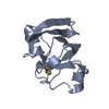 1czpS S: Starting model for refinement C: citing same article ( |
|---|---|
| Similar structure data |


-Links
 PDBj
PDBj


- Assembly
| Deposited unit | 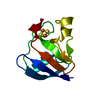
| ||||||||
|---|---|---|---|---|---|---|---|---|---|
| 1 |
| ||||||||
| Unit cell |
|
-Components
| #1: Protein | Mass: 10368.233 Da / Num. of mol.: 1 Source method: isolated from a genetically manipulated source Source: (gene. exp.) |
|---|---|
| #2: Chemical | ChemComp-FES /   Mass: 175.820 Da / Num. of mol.: 1 / Source method: obtained synthetically / Formula: Fe2S2 Mass: 175.820 Da / Num. of mol.: 1 / Source method: obtained synthetically / Formula: Fe2S2 |
| #3: Water | ChemComp-HOH /  Mass: 18.015 Da / Num. of mol.: 115 / Source method: isolated from a natural source / Formula: H2O Mass: 18.015 Da / Num. of mol.: 115 / Source method: isolated from a natural source / Formula: H2O |
| Compound details | FERREDOXINS ARE IRON-SULFUR PROTEINS THAT ARE INVOLVED IN THE TRANSFER OF ELECTRONS IN A WIDE ...FERREDOXIN |
-Experimental details
-Experiment
| Experiment | Method: X-RAY DIFFRACTION / Number of used crystals: 1 |
|---|
- Sample preparation
| Crystal | Density Matthews: 2.34 Å3/Da / Density % sol: 49 % | |||||||||||||||||||||||||||||||||||||||||||||||||
|---|---|---|---|---|---|---|---|---|---|---|---|---|---|---|---|---|---|---|---|---|---|---|---|---|---|---|---|---|---|---|---|---|---|---|---|---|---|---|---|---|---|---|---|---|---|---|---|---|---|---|
| Crystal grow | pH: 6.5 / Details: pH 6.50 | |||||||||||||||||||||||||||||||||||||||||||||||||
| Crystal grow | *PLUS Temperature: 20 ℃ / pH: 7.5 / Method: vapor diffusion, hanging drop | |||||||||||||||||||||||||||||||||||||||||||||||||
| Components of the solutions | *PLUS
|
-Data collection
| Diffraction | Mean temperature: 100 K |
|---|---|
| Diffraction source | Source: ROTATING ANODE / Type: RIGAKU RU200 / Wavelength: 1.5418 |
| Detector | Type: RIGAKU RAXIS IV / Detector: IMAGE PLATE |
| Radiation | Monochromator: MIRRORS / Protocol: SINGLE WAVELENGTH / Monochromatic (M) / Laue (L): M / Scattering type: x-ray |
| Radiation wavelength | Wavelength: 1.5418 Å / Relative weight: 1 |
| Reflection | Resolution: 1.8→30 Å / Num. obs: 8723 / % possible obs: 98.5 % / Redundancy: 7.7 % / Rmerge(I) obs: 0.055 |
| Reflection shell | Resolution: 1.8→1.9 Å / Redundancy: 7.9 % / Rmerge(I) obs: 0.317 / % possible all: 97.2 |
| Reflection | *PLUS Highest resolution: 1.8 Å / Lowest resolution: 33.5 Å / Num. measured all: 161518 / Rmerge(I) obs: 0.055 |
| Reflection shell | *PLUS % possible obs: 97.2 % / Rmerge(I) obs: 0.317 / Mean I/σ(I) obs: 7.7 |
- Processing
| Software |
| ||||||||||||||||||||
|---|---|---|---|---|---|---|---|---|---|---|---|---|---|---|---|---|---|---|---|---|---|
| Refinement | Method to determine structure: MOLECULAR REPLACEMENT Starting model: PDB ENTRY 1CZP Resolution: 1.8→33.52 Å / SU B: 2.689 / SU ML: 0.083 / Cross valid method: THROUGHOUT / ESU R: 0.129 / ESU R Free: 0.125
| ||||||||||||||||||||
| Displacement parameters | Biso mean: 22.385 Å2
| ||||||||||||||||||||
| Refinement step | Cycle: LAST / Resolution: 1.8→33.52 Å
| ||||||||||||||||||||
| Refinement | *PLUS Highest resolution: 1.8 Å / % reflection Rfree: 5 % / Rfactor Rfree: 0.216 / Rfactor Rwork: 0.176 | ||||||||||||||||||||
| Solvent computation | *PLUS | ||||||||||||||||||||
| Displacement parameters | *PLUS | ||||||||||||||||||||
| Refine LS restraints | *PLUS
|
