 Movie
Movie Controller

[English] 日本語
 Yorodumi
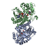
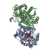
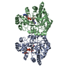
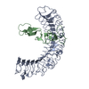
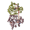
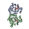
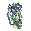
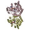
- PDB-1oc2: The structure of NADH in the dTDP-D-glucose dehydratase (RmlB) enzyme -
+ Open data

- Basic information
| Entry | Database: PDB / ID: 1oc2 | ||||||
|---|---|---|---|---|---|---|---|
| Title | The structure of NADH in the dTDP-D-glucose dehydratase (RmlB) enzyme | ||||||
 Components Components | DTDP-GLUCOSE 4,6-DEHYDRATASE | ||||||
 Keywords Keywords | LYASE / DEHYDRATASE / NADH / RHAMNOSE | ||||||
| Function / homology |  Function and homology information Function and homology informationdTDP-glucose 4,6-dehydratase / dTDP-glucose 4,6-dehydratase activity / nucleotide-sugar metabolic process / nucleotide binding Similarity search - Function | ||||||
| Biological species |  STREPTOCOCCUS SUIS (bacteria) STREPTOCOCCUS SUIS (bacteria) | ||||||
| Method |  X-RAY DIFFRACTION / SYNCHROTRON / MOLECULAR REPLACEMENT / Resolution: 1.5 Å X-RAY DIFFRACTION / SYNCHROTRON / MOLECULAR REPLACEMENT / Resolution: 1.5 Å | ||||||
 Authors Authors | Beis, K. / Naismith, J.H. | ||||||
 Citation Citation | Journal: J.Am.Chem.Soc. / Year: 2003 Title: The Structure of Nadh in the Enzyme Dtdp-D-Glucose Dehydratase (Rmlb) Authors: Beis, K. / Allard, S.T. / Hegeman, A.D. / Murshudov, G. / Philp, D. / Naismith, J.H. | ||||||
| History |
|
- Structure visualization
| Structure viewer | Molecule: MolmilJmol/JSmol |
|---|
- Downloads & links
-Download
| PDBx/mmCIF format | 1oc2.cif.gz | 317.8 KB | Display | PDBx/mmCIF format |
|---|---|---|---|---|
| PDB format | pdb1oc2.ent.gz | 257.9 KB | Display | PDB format |
| PDBx/mmJSON format | 1oc2.json.gz | Tree view | PDBx/mmJSON format | |
| Others |  Other downloads Other downloads |
-Validation report
| Arichive directory | https://data.pdbj.org/pub/pdb/validation_reports/oc/1oc2ftp://data.pdbj.org/pub/pdb/validation_reports/oc/1oc2 | HTTPS FTP |
|---|
-Related structure data
| Related structure data | 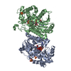 1kepS S: Starting model for refinement |
|---|---|
| Similar structure data |


-Links
 PDBj
PDBj- Assembly
| Deposited unit | 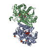
| ||||||||
|---|---|---|---|---|---|---|---|---|---|
| 1 |
| ||||||||
| Unit cell |
|
-Components
| #1: Protein | Mass: 38982.277 Da / Num. of mol.: 2 Source method: isolated from a genetically manipulated source Source: (gene. exp.) STREPTOCOCCUS SUIS (bacteria) / Strain: SEROTYPE 2 / Cell line: BL21DE3 / Plasmid: PET21(+) / Production host: #2: Chemical | 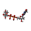  Mass: 534.303 Da / Num. of mol.: 2 / Source method: obtained synthetically / Formula: C15H24N2O15P2 Mass: 534.303 Da / Num. of mol.: 2 / Source method: obtained synthetically / Formula: C15H24N2O15P2#3: Chemical |   Mass: 663.425 Da / Num. of mol.: 2 / Source method: obtained synthetically / Formula: C21H27N7O14P2 / Comment: NAD*YM Mass: 663.425 Da / Num. of mol.: 2 / Source method: obtained synthetically / Formula: C21H27N7O14P2 / Comment: NAD*YM#4: Chemical |   Mass: 96.063 Da / Num. of mol.: 3 / Source method: obtained synthetically / Formula: SO4 Mass: 96.063 Da / Num. of mol.: 3 / Source method: obtained synthetically / Formula: SO4#5: Water | ChemComp-HOH / |  Mass: 18.015 Da / Num. of mol.: 757 / Source method: isolated from a natural source / Formula: H2O Mass: 18.015 Da / Num. of mol.: 757 / Source method: isolated from a natural source / Formula: H2O |
|---|
-Experimental details
-Experiment
| Experiment | Method: X-RAY DIFFRACTION / Number of used crystals: 1 |
|---|
- Sample preparation
| Crystal | Density Matthews: 3.5 Å3/Da / Density % sol: 64.7 % | ||||||||||||||||||||||||||||||||||||||||||||||||
|---|---|---|---|---|---|---|---|---|---|---|---|---|---|---|---|---|---|---|---|---|---|---|---|---|---|---|---|---|---|---|---|---|---|---|---|---|---|---|---|---|---|---|---|---|---|---|---|---|---|
| Crystal grow | pH: 5.4 Details: 30% PEG 4K, 0.1M CITRIC ACID, PH5.4, 0.3M AMMONIUM SULPHATE,, pH 5.40 | ||||||||||||||||||||||||||||||||||||||||||||||||
| Crystal grow | *PLUS pH: 5.4 / Method: vapor diffusion, sitting dropDetails: used microseeding, Allard, S.T., (2002) Structure, 10, 81. | ||||||||||||||||||||||||||||||||||||||||||||||||
| Components of the solutions | *PLUS
|
-Data collection
| Diffraction | Mean temperature: 120 K |
|---|---|
| Diffraction source | Source: SYNCHROTRON / Site: ESRF  / Beamline: ID14-2 / Wavelength: 0.93 / Beamline: ID14-2 / Wavelength: 0.93 |
| Detector | Type: ADSC CCD / Detector: CCD / Date: Nov 15, 2001 / Details: MIRRORS |
| Radiation | Protocol: SINGLE WAVELENGTH / Monochromatic (M) / Laue (L): M / Scattering type: x-ray |
| Radiation wavelength | Wavelength: 0.93 Å / Relative weight: 1 |
| Reflection | Resolution: 1.5→91.28 Å / Num. obs: 179804 / % possible obs: 98.6 % / Redundancy: 3.5 % / Rmerge(I) obs: 0.116 / Net I/σ(I): 4.3 |
| Reflection shell | Resolution: 1.1→1.53 Å / Redundancy: 3.1 % / Rmerge(I) obs: 0.66 / Mean I/σ(I) obs: 1.2 / % possible all: 98.6 |
| Reflection | *PLUS Highest resolution: 1.5 Å / Lowest resolution: 34.7 Å / Num. obs: 168795 / Num. measured all: 2567266 / Rmerge(I) obs: 0.116 |
| Reflection shell | *PLUS Mean I/σ(I) obs: 1.1 |
- Processing
| Software |
| ||||||||||||||||||||||||||||||||||||||||||||||||||||||||||||||||||||||||||||||||||||||||||||||||||||||||||||||||||||||||||||||||||||||||||||||||||||||||||||||||||||||||||||||||||||||
|---|---|---|---|---|---|---|---|---|---|---|---|---|---|---|---|---|---|---|---|---|---|---|---|---|---|---|---|---|---|---|---|---|---|---|---|---|---|---|---|---|---|---|---|---|---|---|---|---|---|---|---|---|---|---|---|---|---|---|---|---|---|---|---|---|---|---|---|---|---|---|---|---|---|---|---|---|---|---|---|---|---|---|---|---|---|---|---|---|---|---|---|---|---|---|---|---|---|---|---|---|---|---|---|---|---|---|---|---|---|---|---|---|---|---|---|---|---|---|---|---|---|---|---|---|---|---|---|---|---|---|---|---|---|---|---|---|---|---|---|---|---|---|---|---|---|---|---|---|---|---|---|---|---|---|---|---|---|---|---|---|---|---|---|---|---|---|---|---|---|---|---|---|---|---|---|---|---|---|---|---|---|---|---|
| Refinement | Method to determine structure: MOLECULAR REPLACEMENT Starting model: PDB ENTRY 1KEP Resolution: 1.5→91.29 Å / Cor.coef. Fo:Fc: 0.966 / Cor.coef. Fo:Fc free: 0.953 / SU B: 1.322 / SU ML: 0.046 / Cross valid method: THROUGHOUT / ESU R: 0.07 / ESU R Free: 0.064 / Stereochemistry target values: MAXIMUM LIKELIHOOD Details: HYDROGENS HAVE BEEN ADDED IN THE RIDING POSITIONS. MISSING RESIDUES FROM REFINEMENT MET A 1 SER A 2 MET B 1 LYS B 348
| ||||||||||||||||||||||||||||||||||||||||||||||||||||||||||||||||||||||||||||||||||||||||||||||||||||||||||||||||||||||||||||||||||||||||||||||||||||||||||||||||||||||||||||||||||||||
| Solvent computation | Ion probe radii: 0.8 Å / Shrinkage radii: 0.8 Å / VDW probe radii: 1.4 Å / Solvent model: BABINET MODEL WITH MASK | ||||||||||||||||||||||||||||||||||||||||||||||||||||||||||||||||||||||||||||||||||||||||||||||||||||||||||||||||||||||||||||||||||||||||||||||||||||||||||||||||||||||||||||||||||||||
| Displacement parameters | Biso mean: 15.63 Å2
| ||||||||||||||||||||||||||||||||||||||||||||||||||||||||||||||||||||||||||||||||||||||||||||||||||||||||||||||||||||||||||||||||||||||||||||||||||||||||||||||||||||||||||||||||||||||
| Refinement step | Cycle: LAST / Resolution: 1.5→91.29 Å
| ||||||||||||||||||||||||||||||||||||||||||||||||||||||||||||||||||||||||||||||||||||||||||||||||||||||||||||||||||||||||||||||||||||||||||||||||||||||||||||||||||||||||||||||||||||||
| Refine LS restraints |
|