 Movie
Movie Controller


+ Open data

- Basic information
| Entry | Database: PDB / ID: 1obd | ||||||
|---|---|---|---|---|---|---|---|
| Title | SAICAR-synthase complexed with ATP | ||||||
 Components Components | PHOSPHORIBOSYLAMIDOIMIDAZOLE- SUCCINOCARBOXAMIDE SYNTHASE | ||||||
 Keywords Keywords | LIGASE / SYNTHETASE / ATP BINDING PROTEIN / PHOSPHORIBOSYLAMINOIMIDAZOLESUCCINOCARBOXAMIDE (SAICAR) SYNTHASE / PURINE BIOSYNTHESIS | ||||||
| Function / homology |  Function and homology information Function and homology informationphosphoribosylaminoimidazolesuccinocarboxamide synthase / phosphoribosylaminoimidazolesuccinocarboxamide synthase activity / purine nucleotide biosynthetic process / 'de novo' IMP biosynthetic process / ATP binding / nucleus / cytoplasm Similarity search - Function | ||||||
| Biological species |  | ||||||
| Method |  X-RAY DIFFRACTION / SYNCHROTRON / MOLECULAR REPLACEMENT / Resolution: 1.4 Å X-RAY DIFFRACTION / SYNCHROTRON / MOLECULAR REPLACEMENT / Resolution: 1.4 Å | ||||||
 Authors Authors | Antonyuk, S.V. / Grebenko, A.I. / Levdikov, V.M. / Urusova, D.V. / Melik-Adamyan, W.R. / Lamzin, V.S. / Wilson, K. | ||||||
 Citation Citation | Journal: Kristallografiya / Year: 2001 Title: X-Ray Structure of Saicar-Synthase Complexed with ATP Authors: Antonyuk, S.V. / Grebenko, A.I. / Levdikov, V.M. / Urusova, D.V. / Melik-Adamyan, W.R. / Lamzin, V.S. / Wilson, K. #1: Journal: Structure / Year: 1998Title: The Structure of Saicar Synthase: An Enzyme in the De Novo Pathway of Purine Nucleotide Biosynthesis Authors: Levdikov, V.M. / Barynin, V.V. / Grebenko, A.I. / Melik-Adamyan, W.R. / Lamzin, V.S. / Wilson, K.S. | ||||||
| History |
| ||||||
| Remark 700 | SHEET THE SHEET STRUCTURE OF THIS MOLECULE IS BIFURCATED. IN ORDER TO REPRESENT THIS FEATURE IN ... SHEET THE SHEET STRUCTURE OF THIS MOLECULE IS BIFURCATED. IN ORDER TO REPRESENT THIS FEATURE IN THE SHEET RECORDS BELOW, TWO SHEETS ARE DEFINED. |
- Structure visualization
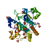
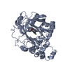
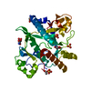
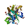
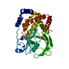
| Structure viewer | Molecule: MolmilJmol/JSmol |
|---|
- Downloads & links
-Download
| PDBx/mmCIF format | 1obd.cif.gz | 161.9 KB | Display | PDBx/mmCIF format |
|---|---|---|---|---|
| PDB format | pdb1obd.ent.gz | 126.2 KB | Display | PDB format |
| PDBx/mmJSON format | 1obd.json.gz | Tree view | PDBx/mmJSON format | |
| Others |  Other downloads Other downloads |
-Validation report
| Arichive directory | https://data.pdbj.org/pub/pdb/validation_reports/ob/1obdftp://data.pdbj.org/pub/pdb/validation_reports/ob/1obd | HTTPS FTP |
|---|
-Related structure data
| Related structure data | 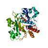 1obgC  1a48S S: Starting model for refinement C: citing same article ( |
|---|---|
| Similar structure data |


-Links
 PDBj
PDBj


- Assembly
| Deposited unit | 
| ||||||||
|---|---|---|---|---|---|---|---|---|---|
| 1 |
| ||||||||
| Unit cell |
|
-Components
-Protein , 1 types, 1 molecules A
| #1: Protein | Mass: 34545.238 Da / Num. of mol.: 1 / Source method: isolated from a natural source / Source: (natural) References: UniProt: P27616, phosphoribosylaminoimidazolesuccinocarboxamide synthase |
|---|
-Non-polymers , 5 types, 532 molecules 


| #2: Chemical | ChemComp-ATP /  Mass: 507.181 Da / Num. of mol.: 1 / Source method: obtained synthetically / Formula: C10H16N5O13P3 / Comment: ATP, energy-carrying molecule*YM Mass: 507.181 Da / Num. of mol.: 1 / Source method: obtained synthetically / Formula: C10H16N5O13P3 / Comment: ATP, energy-carrying molecule*YM | ||||||
|---|---|---|---|---|---|---|---|
| #3: Chemical |  Mass: 96.063 Da / Num. of mol.: 2 / Source method: obtained synthetically / Formula: SO4 Mass: 96.063 Da / Num. of mol.: 2 / Source method: obtained synthetically / Formula: SO4#4: Chemical |  Mass: 24.305 Da / Num. of mol.: 2 / Source method: obtained synthetically / Formula: Mg Mass: 24.305 Da / Num. of mol.: 2 / Source method: obtained synthetically / Formula: Mg#5: Chemical | ChemComp-AMP / |  Mass: 347.221 Da / Num. of mol.: 1 / Source method: obtained synthetically / Formula: C10H14N5O7P / Comment: AMP*YM Mass: 347.221 Da / Num. of mol.: 1 / Source method: obtained synthetically / Formula: C10H14N5O7P / Comment: AMP*YM#6: Water | ChemComp-HOH / | Mass: 18.015 Da / Num. of mol.: 526 / Source method: isolated from a natural source / Formula: H2O |
-Details
| Has protein modification | Y |
|---|
-Experimental details
-Experiment
| Experiment | Method: X-RAY DIFFRACTION / Number of used crystals: 1 |
|---|
- Sample preparation
| Crystal | Density Matthews: 2.2 Å3/Da / Density % sol: 44.8 % |
|---|---|
| Crystal grow | Method: vapor diffusion, hanging drop / pH: 7.5 Details: THE HANGING DROPS CONTAINED PROTEIN AT A CONCENTRATION OF 8 - 15 MG/ML, 50 MM TRIS-HCL BUFFER, PH 7.0, 40 MM ASPARTIC ACID AND 1.0 - 1.25 M AMMONIUM SULPHATE. THE WELL CONTAINED THE SAME ...Details: THE HANGING DROPS CONTAINED PROTEIN AT A CONCENTRATION OF 8 - 15 MG/ML, 50 MM TRIS-HCL BUFFER, PH 7.0, 40 MM ASPARTIC ACID AND 1.0 - 1.25 M AMMONIUM SULPHATE. THE WELL CONTAINED THE SAME ABOVE BUFFER PLUS 2.25 M AMMONIUM SULPHATE |
| Crystal grow | *PLUS Method: unknown |
-Data collection
| Diffraction | Mean temperature: 100 K |
|---|---|
| Diffraction source | Source: SYNCHROTRON / Site: EMBL/DESY, HAMBURG  / Beamline: X11 / Wavelength: 0.906 / Beamline: X11 / Wavelength: 0.906 |
| Detector | Type: MARRESEARCH / Detector: IMAGE PLATE / Date: Feb 15, 1998 |
| Radiation | Protocol: SINGLE WAVELENGTH / Monochromatic (M) / Laue (L): M / Scattering type: x-ray |
| Radiation wavelength | Wavelength: 0.906 Å / Relative weight: 1 |
| Reflection | Resolution: 1.4→30 Å / Num. obs: 59985 / % possible obs: 97 % / Redundancy: 3.5 % / Biso Wilson estimate: 16.6 Å2 / Rmerge(I) obs: 0.085 / Net I/σ(I): 17 |
| Reflection shell | Resolution: 1.4→1.42 Å / Redundancy: 3.5 % / Rmerge(I) obs: 0.19 / Mean I/σ(I) obs: 7 / % possible all: 97.4 |
| Reflection shell | *PLUS % possible obs: 97.4 % |
- Processing
| Software |
| ||||||||||||||||||||||||||||||||||||
|---|---|---|---|---|---|---|---|---|---|---|---|---|---|---|---|---|---|---|---|---|---|---|---|---|---|---|---|---|---|---|---|---|---|---|---|---|---|
| Refinement | Method to determine structure: MOLECULAR REPLACEMENT Starting model: PDB ENTRY 1A48 Resolution: 1.4→30 Å / SU B: 0.775 / SU ML: 0.033 / Cross valid method: THROUGHOUT / ESU R: 0.077 / ESU R Free: 0.069 / Details: HYDROGENS HAVE BEEN ADDED IN THE RIDING POSITIONS
| ||||||||||||||||||||||||||||||||||||
| Displacement parameters | Biso mean: 15.9 Å2
| ||||||||||||||||||||||||||||||||||||
| Refinement step | Cycle: LAST / Resolution: 1.4→30 Å
| ||||||||||||||||||||||||||||||||||||
| Software | *PLUS Name: REFMAC / Classification: refinement | ||||||||||||||||||||||||||||||||||||
| Refine LS restraints | *PLUS
|
