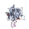
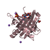
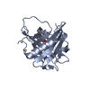
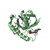
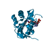
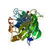
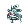
- PDB-1o3y: Crystal structure of mouse ARF1 (delta17-Q71L), GTP form -
+
Open data
ID or keywords:
Loading...
-
Basic information
Entry
Database: PDB / ID: 1o3y
Title
Crystal structure of mouse ARF1 (delta17-Q71L), GTP form
Components
ADP-ribosylation factor 1
Keywords
PROTEIN TRANSPORT
Function / homology
Function and homology information
Glycosphingolipid transport / synaptic vesicle budding / positive regulation of late endosome to lysosome transport / : / trans-Golgi Network Vesicle Budding / lysosomal membrane organization / regulation of phospholipid metabolic process / phospholipase D activator activity / Synthesis of PIPs at the Golgi membrane / Intra-Golgi traffic ...Glycosphingolipid transport / synaptic vesicle budding / positive regulation of late endosome to lysosome transport / : / trans-Golgi Network Vesicle Budding / lysosomal membrane organization / regulation of phospholipid metabolic process / phospholipase D activator activity / Synthesis of PIPs at the Golgi membrane / Intra-Golgi traffic / COPI-coated vesicle / very-low-density lipoprotein particle assembly / Lysosome Vesicle Biogenesis / postsynaptic actin cytoskeleton organization / COPI-mediated anterograde transport / mitotic cleavage furrow ingression / Synthesis of PIPs at the plasma membrane / positive regulation of ER to Golgi vesicle-mediated transport / regulation of receptor internalization / COPI-dependent Golgi-to-ER retrograde traffic / Golgi Associated Vesicle Biogenesis / regulation of Arp2/3 complex-mediated actin nucleation / positive regulation of calcium ion-dependent exocytosis / MHC class II antigen presentation / dendritic spine organization / long-term synaptic depression / peroxisomal membrane / positive regulation of sodium ion transmembrane transport / positive regulation of dendritic spine development / cell leading edge / intracellular copper ion homeostasis / positive regulation of endocytosis / vesicle-mediated transport / endomembrane system / actin filament organization / positive regulation of protein secretion / small monomeric GTPase / sarcomere / trans-Golgi network / intracellular protein transport / cellular response to virus / GDP binding / late endosome / protein transport / G protein activity / neuron projection / postsynaptic density / Golgi membrane / protein domain specific binding / GTP binding / glutamatergic synapse / Golgi apparatus / magnesium ion binding / protein-containing complex / plasma membrane / cytosol / cytoplasm Similarity search - Function
ADP-ribosylation factor 1-5 / Small GTPase superfamily, ARF type / Small GTPase Arf domain profile. / Sar1p-like members of the Ras-family of small GTPases / Small GTPase superfamily, ARF/SAR type / ADP-ribosylation factor family / ARF-like small GTPases; ARF, ADP-ribosylation factor / Rab subfamily of small GTPases / Small GTP-binding protein domain / P-loop containing nucleotide triphosphate hydrolases ...ADP-ribosylation factor 1-5 / Small GTPase superfamily, ARF type / Small GTPase Arf domain profile. / Sar1p-like members of the Ras-family of small GTPases / Small GTPase superfamily, ARF/SAR type / ADP-ribosylation factor family / ARF-like small GTPases; ARF, ADP-ribosylation factor / Rab subfamily of small GTPases / Small GTP-binding protein domain / P-loop containing nucleotide triphosphate hydrolases / Rossmann fold / P-loop containing nucleoside triphosphate hydrolase / 3-Layer(aba) Sandwich / Alpha Beta Similarity search - Domain/homology
In the structure databanks used in Yorodumi, some data are registered as the other names, "COVID-19 virus" and "2019-nCoV". Here are the details of the virus and the list of structure data.
Jan 31, 2019. EMDB accession codes are about to change! (news from PDBe EMDB page)
EMDB accession codes are about to change! (news from PDBe EMDB page)
The allocation of 4 digits for EMDB accession codes will soon come to an end. Whilst these codes will remain in use, new EMDB accession codes will include an additional digit and will expand incrementally as the available range of codes is exhausted. The current 4-digit format prefixed with “EMD-” (i.e. EMD-XXXX) will advance to a 5-digit format (i.e. EMD-XXXXX), and so on. It is currently estimated that the 4-digit codes will be depleted around Spring 2019, at which point the 5-digit format will come into force.
The EM Navigator/Yorodumi systems omit the EMD- prefix.
Related info.:Q: What is EMD? / ID/Accession-code notation in Yorodumi/EM Navigator
Yorodumi is a browser for structure data from EMDB, PDB, SASBDB, etc.
This page is also the successor to EM Navigator detail page, and also detail information page/front-end page for Omokage search.
The word "yorodu" (or yorozu) is an old Japanese word meaning "ten thousand". "mi" (miru) is to see.
Related info.:EMDB / PDB / SASBDB / Comparison of 3 databanks / Yorodumi Search / Aug 31, 2016. New EM Navigator & Yorodumi / Yorodumi Papers / Jmol/JSmol / Function and homology information / Changes in new EM Navigator and Yorodumi
 Movie
Movie Controller


 Open data
 Basic information
 Components
 Keywords
 Function and homology information
 X-RAY DIFFRACTION /
  Authors
 Citation
 Structure visualization
 Downloads & links
 Other downloads
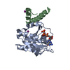


 PDBj
PDBj


 Assembly
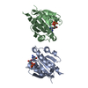
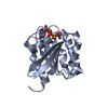


 Mass: 24.305 Da / Num. of mol.: 2 / Source method: obtained synthetically / Formula: Mg
 Mass: 523.180 Da / Num. of mol.: 2 / Source method: obtained synthetically / Formula: C10H16N5O14P3 / Comment: GTP, energy-carrying molecule*YM
 Mass: 18.015 Da / Num. of mol.: 313 / Source method: isolated from a natural source / Formula: H2O
 Sample preparation
 / Beamline: BL-6A / Wavelength: 0.977 Å
 Processing