 Movie
Movie Controller

[English] 日本語
 Yorodumi
- PDB-1nyr: Structure of Staphylococcus aureus threonyl-tRNA synthetase compl... -
+ Open data

- Basic information
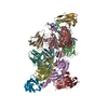
| Entry | Database: PDB / ID: 1nyr | ||||||
|---|---|---|---|---|---|---|---|
| Title | Structure of Staphylococcus aureus threonyl-tRNA synthetase complexed with ATP | ||||||
 Components Components | threonyl-tRNA synthetase 1 | ||||||
 Keywords Keywords | LIGASE / threonyl-tRNA synthetase / ATP / threonine | ||||||
| Function / homology |  Function and homology information Function and homology informationcatalytic activity, acting on a protein / threonine-tRNA ligase / threonyl-tRNA aminoacylation / threonine-tRNA ligase activity / transferase activity / tRNA binding / ATP binding / metal ion binding / cytoplasm Similarity search - Function | ||||||
| Biological species |   Staphylococcus aureus (bacteria) Staphylococcus aureus (bacteria) | ||||||
| Method |  X-RAY DIFFRACTION / SYNCHROTRON / MOLECULAR REPLACEMENT / Resolution: 2.8 Å X-RAY DIFFRACTION / SYNCHROTRON / MOLECULAR REPLACEMENT / Resolution: 2.8 Å | ||||||
 Authors Authors | Torres-Larios, A. / Sankaranarayanan, R. / Rees, B. / Dock-Bregeon, A.C. / Moras, D. | ||||||
 Citation Citation | Journal: J.Mol.Biol. / Year: 2003 Title: Conformational movements and cooperativity upon amino acid, ATP and tRNA binding in threonyl-tRNA synthetase Authors: Torres-Larios, A. / Sankaranarayanan, R. / Rees, B. / Dock-Bregeon, A.C. / Moras, D. | ||||||
| History |
|
- Structure visualization
| Structure viewer | Molecule: MolmilJmol/JSmol |
|---|
- Downloads & links
-Download
| PDBx/mmCIF format | 1nyr.cif.gz | 274.6 KB | Display | PDBx/mmCIF format |
|---|---|---|---|---|
| PDB format | pdb1nyr.ent.gz | 219.2 KB | Display | PDB format |
| PDBx/mmJSON format | 1nyr.json.gz | Tree view | PDBx/mmJSON format | |
| Others |  Other downloads Other downloads |
-Validation report
| Arichive directory | https://data.pdbj.org/pub/pdb/validation_reports/ny/1nyrftp://data.pdbj.org/pub/pdb/validation_reports/ny/1nyr | HTTPS FTP |
|---|
-Related structure data
| Related structure data |  1nyqC  1qf6S S: Starting model for refinement C: citing same article ( |
|---|---|
| Similar structure data |


-Links
 PDBj
PDBj


- Assembly
| Deposited unit | 
| ||||||||
|---|---|---|---|---|---|---|---|---|---|
| 1 |
| ||||||||
| Unit cell |
| ||||||||
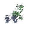
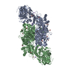
| Details | The protein is a homodimer made of chains A and B |
-Components
| #1: Protein | Mass: 74562.156 Da / Num. of mol.: 2 Source method: isolated from a genetically manipulated source Source: (gene. exp.) Staphylococcus aureus (bacteria) / Plasmid: pBlue-Pet / Species (production host): Escherichia coli / Production host: #2: Chemical |   Mass: 65.409 Da / Num. of mol.: 3 / Source method: obtained synthetically / Formula: Zn Mass: 65.409 Da / Num. of mol.: 3 / Source method: obtained synthetically / Formula: Zn#3: Chemical |   Mass: 507.181 Da / Num. of mol.: 2 / Source method: obtained synthetically / Formula: C10H16N5O13P3 / Comment: ATP, energy-carrying molecule*YM Mass: 507.181 Da / Num. of mol.: 2 / Source method: obtained synthetically / Formula: C10H16N5O13P3 / Comment: ATP, energy-carrying molecule*YM#4: Chemical | 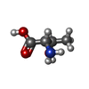  Type: L-peptide linking / Mass: 119.119 Da / Num. of mol.: 2 / Source method: obtained synthetically / Formula: C4H9NO3 Type: L-peptide linking / Mass: 119.119 Da / Num. of mol.: 2 / Source method: obtained synthetically / Formula: C4H9NO3#5: Water | ChemComp-HOH / |  Mass: 18.015 Da / Num. of mol.: 216 / Source method: isolated from a natural source / Formula: H2O Mass: 18.015 Da / Num. of mol.: 216 / Source method: isolated from a natural source / Formula: H2O |
|---|
-Experimental details
-Experiment
| Experiment | Method: X-RAY DIFFRACTION / Number of used crystals: 1 |
|---|
- Sample preparation
| Crystal | Density Matthews: 3.17 Å3/Da / Density % sol: 60.89 % | ||||||||||||||||||||||||||||||||||||||||||||||||||||||||
|---|---|---|---|---|---|---|---|---|---|---|---|---|---|---|---|---|---|---|---|---|---|---|---|---|---|---|---|---|---|---|---|---|---|---|---|---|---|---|---|---|---|---|---|---|---|---|---|---|---|---|---|---|---|---|---|---|---|
| Crystal grow | Temperature: 277 K / Method: vapor diffusion, hanging drop / pH: 8 Details: PEG 8000, Tris.Cl, potassium chloride, VAPOR DIFFUSION, HANGING DROP, temperature 277K | ||||||||||||||||||||||||||||||||||||||||||||||||||||||||
| Crystal grow | *PLUS Temperature: 4 ℃ / pH: 7.2 / Method: vapor diffusion, hanging drop | ||||||||||||||||||||||||||||||||||||||||||||||||||||||||
| Components of the solutions | *PLUS
|
-Data collection
| Diffraction | Mean temperature: 100 K |
|---|---|
| Diffraction source | Source: SYNCHROTRON / Site: ESRF  / Beamline: BM30A / Wavelength: 0.9797 Å / Beamline: BM30A / Wavelength: 0.9797 Å |
| Detector | Type: MARRESEARCH / Detector: CCD / Date: Nov 22, 1999 / Details: mirrors |
| Radiation | Monochromator: 2 Silicon crystals / Protocol: SINGLE WAVELENGTH / Monochromatic (M) / Laue (L): M / Scattering type: x-ray |
| Radiation wavelength | Wavelength: 0.9797 Å / Relative weight: 1 |
| Reflection | Resolution: 2.8→25 Å / Num. all: 46973 / Num. obs: 46973 / % possible obs: 98.3 % / Observed criterion σ(F): 0 / Redundancy: 3.4 % / Biso Wilson estimate: 79.9 Å2 / Rmerge(I) obs: 0.036 / Net I/σ(I): 19.1 |
| Reflection shell | Resolution: 2.8→2.87 Å / Rmerge(I) obs: 0.29 / Mean I/σ(I) obs: 3.8 / Num. unique all: 2998 / % possible all: 95.7 |
| Reflection | *PLUS Num. measured all: 160435 |
| Reflection shell | *PLUS Highest resolution: 2.8 Å / % possible obs: 95.7 % / Num. unique obs: 2998 / Rmerge(I) obs: 0.29 |
- Processing
| Software |
| ||||||||||||||||||||||||||||||||||||
|---|---|---|---|---|---|---|---|---|---|---|---|---|---|---|---|---|---|---|---|---|---|---|---|---|---|---|---|---|---|---|---|---|---|---|---|---|---|
| Refinement | Method to determine structure: MOLECULAR REPLACEMENT Starting model: PDB entry 1QF6 Resolution: 2.8→15 Å / Rfactor Rfree error: 0.007 / Isotropic thermal model: RESTRAINED / Cross valid method: THROUGHOUT / σ(F): 0 / Stereochemistry target values: Engh & Huber
| ||||||||||||||||||||||||||||||||||||
| Solvent computation | Solvent model: FLAT MODEL / Bsol: 58.1117 Å2 / ksol: 0.334807 e/Å3 | ||||||||||||||||||||||||||||||||||||
| Displacement parameters | Biso mean: 73.6 Å2
| ||||||||||||||||||||||||||||||||||||
| Refine analyze |
| ||||||||||||||||||||||||||||||||||||
| Refinement step | Cycle: LAST / Resolution: 2.8→15 Å
| ||||||||||||||||||||||||||||||||||||
| Refine LS restraints |
| ||||||||||||||||||||||||||||||||||||
| LS refinement shell | Resolution: 2.8→2.97 Å / Rfactor Rfree error: 0.02 / Total num. of bins used: 6
| ||||||||||||||||||||||||||||||||||||
| Xplor file |
| ||||||||||||||||||||||||||||||||||||
| Refinement | *PLUS Highest resolution: 2.8 Å / Lowest resolution: 15 Å | ||||||||||||||||||||||||||||||||||||
| Solvent computation | *PLUS | ||||||||||||||||||||||||||||||||||||
| Displacement parameters | *PLUS | ||||||||||||||||||||||||||||||||||||
| Refine LS restraints | *PLUS
|
