 Movie
Movie Controller

[English] 日本語
 Yorodumi
- PDB-1nvw: Structural evidence for feedback activation by RasGTP of the Ras-... -
+ Open data

- Basic information
| Entry | Database: PDB / ID: 1nvw | ||||||
|---|---|---|---|---|---|---|---|
| Title | Structural evidence for feedback activation by RasGTP of the Ras-specific nucleotide exchange factor SOS | ||||||
 Components Components |
| ||||||
 Keywords Keywords | SIGNALING PROTEIN / Proto-oncogene / GTP-binding / Guanine-nucleotide releasing factor | ||||||
| Function / homology |  Function and homology information Function and homology informationGTPase complex / phospholipase C activator activity / Interleukin-15 signaling / oncogene-induced cell senescence / Activation of RAC1 / positive regulation of miRNA metabolic process / Signaling by LTK / leukocyte migration / T-helper 1 type immune response / epidermal growth factor receptor binding ...GTPase complex / phospholipase C activator activity / Interleukin-15 signaling / oncogene-induced cell senescence / Activation of RAC1 / positive regulation of miRNA metabolic process / Signaling by LTK / leukocyte migration / T-helper 1 type immune response / epidermal growth factor receptor binding / Regulation of KIT signaling / neurotrophin TRK receptor signaling pathway / NRAGE signals death through JNK / positive regulation of ruffle assembly / defense response to protozoan / positive regulation of wound healing / Fc-epsilon receptor signaling pathway / GRB2:SOS provides linkage to MAPK signaling for Integrins / regulation of neurotransmitter receptor localization to postsynaptic specialization membrane / adipose tissue development / RET signaling / Signaling by RAS GAP mutants / Signaling by RAS GTPase mutants / Activation of RAS in B cells / RAS signaling downstream of NF1 loss-of-function variants / Role of LAT2/NTAL/LAB on calcium mobilization / Interleukin receptor SHC signaling / SOS-mediated signalling / Signal attenuation / positive regulation of protein targeting to membrane / Activated NTRK3 signals through RAS / Activated NTRK2 signals through RAS / SHC1 events in ERBB4 signaling / Signalling to RAS / Schwann cell development / SHC-related events triggered by IGF1R / Activated NTRK2 signals through FRS2 and FRS3 / Estrogen-stimulated signaling through PRKCZ / positive regulation of Rac protein signal transduction / SHC-mediated cascade:FGFR3 / positive regulation of epidermal growth factor receptor signaling pathway / MET activates RAS signaling / SHC-mediated cascade:FGFR2 / SHC-mediated cascade:FGFR4 / Signaling by PDGFRA transmembrane, juxtamembrane and kinase domain mutants / Signaling by PDGFRA extracellular domain mutants / PTK6 Regulates RHO GTPases, RAS GTPase and MAP kinases / Erythropoietin activates RAS / SHC-mediated cascade:FGFR1 / Signaling by FGFR4 in disease / FRS-mediated FGFR3 signaling / Signaling by FLT3 ITD and TKD mutants / FRS-mediated FGFR2 signaling / FRS-mediated FGFR4 signaling / p38MAPK events / FRS-mediated FGFR1 signaling / Signaling by FGFR3 in disease / RAC1 GTPase cycle / Tie2 Signaling / protein-membrane adaptor activity / Signaling by FGFR2 in disease / myelination / EPHB-mediated forward signaling / insulin-like growth factor receptor signaling pathway / Signaling by FLT3 fusion proteins / GRB2 events in EGFR signaling / SHC1 events in EGFR signaling / FLT3 Signaling / Signaling by FGFR1 in disease / EGFR Transactivation by Gastrin / FCERI mediated Ca+2 mobilization / NCAM signaling for neurite out-growth / CD209 (DC-SIGN) signaling / positive regulation of epithelial cell proliferation / GRB2 events in ERBB2 signaling / Downstream signal transduction / intrinsic apoptotic signaling pathway / Insulin receptor signalling cascade / guanyl-nucleotide exchange factor activity / SHC1 events in ERBB2 signaling / response to ischemia / Constitutive Signaling by Overexpressed ERBB2 / Ras activation upon Ca2+ influx through NMDA receptor / Antigen activates B Cell Receptor (BCR) leading to generation of second messengers / Signaling by phosphorylated juxtamembrane, extracellular and kinase domain KIT mutants / axon guidance / animal organ morphogenesis / B cell receptor signaling pathway / GTPase activator activity / VEGFR2 mediated cell proliferation / T cell activation / regulation of actin cytoskeleton organization / small monomeric GTPase / FCERI mediated MAPK activation / regulation of long-term neuronal synaptic plasticity / molecular condensate scaffold activity / Signaling by ERBB2 TMD/JMD mutants / Signaling by SCF-KIT / RAF activation / Constitutive Signaling by EGFRvIII Similarity search - Function | ||||||
| Biological species |  Homo sapiens (human) Homo sapiens (human) | ||||||
| Method |  X-RAY DIFFRACTION / SYNCHROTRON / FOURIER SYNTHESIS / Resolution: 2.7 Å X-RAY DIFFRACTION / SYNCHROTRON / FOURIER SYNTHESIS / Resolution: 2.7 Å | ||||||
 Authors Authors | Margarit, S.M. / Sondermann, H. / Hall, B.E. / Nagar, B. / Hoelz, A. / Pirruccello, M. / Bar-Sagi, D. / Kuriyan, J. | ||||||
 Citation Citation | Journal: Cell(Cambridge,Mass.) / Year: 2003 Title: Structural evidence for feedback activation by RasGTP of the Ras-specific nucleotide exchange factor SOS Authors: Margarit, S.M. / Sondermann, H. / Hall, B.E. / Nagar, B. / Hoelz, A. / Pirruccello, M. / Bar-Sagi, D. / Kuriyan, J. | ||||||
| History |
|
- Structure visualization
| Structure viewer | Molecule: MolmilJmol/JSmol |
|---|
- Downloads & links
-Download
| PDBx/mmCIF format | 1nvw.cif.gz | 179.1 KB | Display | PDBx/mmCIF format |
|---|---|---|---|---|
| PDB format | pdb1nvw.ent.gz | 138.9 KB | Display | PDB format |
| PDBx/mmJSON format | 1nvw.json.gz | Tree view | PDBx/mmJSON format | |
| Others |  Other downloads Other downloads |
-Validation report
| Arichive directory | https://data.pdbj.org/pub/pdb/validation_reports/nv/1nvwftp://data.pdbj.org/pub/pdb/validation_reports/nv/1nvw | HTTPS FTP |
|---|
-Related structure data
| Related structure data | 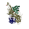 1nvuSC 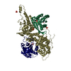 1nvvC 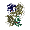 1nvxC S: Starting model for refinement C: citing same article ( |
|---|---|
| Similar structure data |


-Links
 PDBj
PDBj


- Assembly
| Deposited unit | 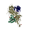
| ||||||||
|---|---|---|---|---|---|---|---|---|---|
| 1 |
| ||||||||
| 2 | x 8
| ||||||||
| 3 | 
| ||||||||
| Unit cell |
| ||||||||
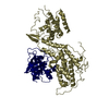
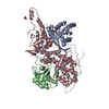
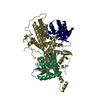
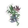
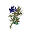
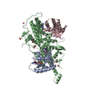
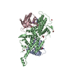
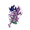
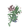
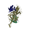
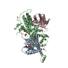
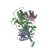
| Details | Two molecules of Ras bound to one molecule of Sos, one at the active site (chainID, R) and the other at a distal site (chainID, Q). The distal Ras has GTP bound to it, while the active site Ras is nucleotide-free. |
-Components
-Protein , 2 types, 3 molecules QRS
| #1: Protein | Mass: 18875.191 Da / Num. of mol.: 2 / Fragment: RESIDUES 1-166 Source method: isolated from a genetically manipulated source Source: (gene. exp.) Homo sapiens (human) / Gene: HRAS OR HRAS1 / Plasmid: p-ProEXHT / Species (production host): Escherichia coli / Production host:  #2: Protein | | Mass: 56532.730 Da / Num. of mol.: 1 Fragment: RESIDUES 566-1046, INCLUDING RAS GUANINE NUCLEOTIDE EXCHANGE FACTOR FRAGMENT Source method: isolated from a genetically manipulated source Source: (gene. exp.) Homo sapiens (human) / Gene: SOS1 / Plasmid: p-ProEXHT / Species (production host): Escherichia coli / Production host: |
|---|
-Non-polymers , 4 types, 64 molecules 


| #3: Chemical | ChemComp-MG /  Mass: 24.305 Da / Num. of mol.: 1 / Source method: obtained synthetically / Formula: Mg Mass: 24.305 Da / Num. of mol.: 1 / Source method: obtained synthetically / Formula: Mg | ||||
|---|---|---|---|---|---|
| #4: Chemical | ChemComp-PO4 /  Mass: 94.971 Da / Num. of mol.: 7 / Source method: obtained synthetically / Formula: PO4 Mass: 94.971 Da / Num. of mol.: 7 / Source method: obtained synthetically / Formula: PO4#5: Chemical | ChemComp-GNP / |  Mass: 522.196 Da / Num. of mol.: 1 / Source method: obtained synthetically / Formula: C10H17N6O13P3 Mass: 522.196 Da / Num. of mol.: 1 / Source method: obtained synthetically / Formula: C10H17N6O13P3Comment: GppNHp, GMPPNP, energy-carrying molecule analogue*YM #6: Water | ChemComp-HOH / | Mass: 18.015 Da / Num. of mol.: 55 / Source method: isolated from a natural source / Formula: H2O |
-Experimental details
-Experiment
| Experiment | Method: X-RAY DIFFRACTION / Number of used crystals: 1 |
|---|
- Sample preparation
| Crystal | Density Matthews: 4.06 Å3/Da / Density % sol: 69.47 % | ||||||||||||||||||||||||
|---|---|---|---|---|---|---|---|---|---|---|---|---|---|---|---|---|---|---|---|---|---|---|---|---|---|
| Crystal grow | Temperature: 277 K / Method: vapor diffusion, hanging drop / pH: 7.5 Details: 1.2-1.5 M phosphate, 100 mM Hepes, pH 7.5, VAPOR DIFFUSION, HANGING DROP, temperature 277K | ||||||||||||||||||||||||
| Crystal grow | *PLUS | ||||||||||||||||||||||||
| Components of the solutions | *PLUS
|
-Data collection
| Diffraction | Mean temperature: 100 K |
|---|---|
| Diffraction source | Source: SYNCHROTRON / Site: ALS  / Beamline: 8.2.2 / Wavelength: 1 Å / Beamline: 8.2.2 / Wavelength: 1 Å |
| Detector | Type: ADSC QUANTUM 315 / Detector: CCD / Date: Nov 25, 2002 / Details: mirrors |
| Radiation | Monochromator: Double crystal Si(111) / Protocol: SINGLE WAVELENGTH / Monochromatic (M) / Laue (L): M / Scattering type: x-ray |
| Radiation wavelength | Wavelength: 1 Å / Relative weight: 1 |
| Reflection | Resolution: 2.7→50 Å / Num. all: 42132 / Num. obs: 42132 / % possible obs: 100 % / Observed criterion σ(F): 0 / Observed criterion σ(I): -3 / Redundancy: 7.7 % / Biso Wilson estimate: 56.9 Å2 / Rsym value: 0.101 / Net I/σ(I): 20 |
| Reflection shell | Resolution: 2.7→2.8 Å / Redundancy: 7.6 % / Mean I/σ(I) obs: 4.6 / Num. unique all: 4146 / Rsym value: 0.603 / % possible all: 100 |
| Reflection | *PLUS Highest resolution: 2.7 Å / Lowest resolution: 64 Å / Num. obs: 40362 / % possible obs: 99.1 % / Num. measured all: 313273 / Rmerge(I) obs: 0.088 |
| Reflection shell | *PLUS % possible obs: 96.7 % / Rmerge(I) obs: 0.463 / Mean I/σ(I) obs: 2.6 |
- Processing
| Software |
| ||||||||||||||||||||||||||||||||||||
|---|---|---|---|---|---|---|---|---|---|---|---|---|---|---|---|---|---|---|---|---|---|---|---|---|---|---|---|---|---|---|---|---|---|---|---|---|---|
| Refinement | Method to determine structure: FOURIER SYNTHESIS Starting model: PDB ENTRY 1NVU Resolution: 2.7→49.09 Å / Rfactor Rfree error: 0.005 / Isotropic thermal model: RESTRAINED / Cross valid method: THROUGHOUT / σ(F): 0 / σ(I): 0 / Stereochemistry target values: Engh & Huber
| ||||||||||||||||||||||||||||||||||||
| Solvent computation | Solvent model: FLAT MODEL / Bsol: 21.2438 Å2 / ksol: 0.347129 e/Å3 | ||||||||||||||||||||||||||||||||||||
| Displacement parameters | Biso mean: 45 Å2
| ||||||||||||||||||||||||||||||||||||
| Refine analyze |
| ||||||||||||||||||||||||||||||||||||
| Refinement step | Cycle: LAST / Resolution: 2.7→49.09 Å
| ||||||||||||||||||||||||||||||||||||
| Refine LS restraints |
| ||||||||||||||||||||||||||||||||||||
| LS refinement shell | Resolution: 2.7→2.87 Å / Rfactor Rfree error: 0.016 / Total num. of bins used: 6
| ||||||||||||||||||||||||||||||||||||
| Xplor file |
| ||||||||||||||||||||||||||||||||||||
| Refinement | *PLUS Lowest resolution: 64 Å / Num. reflection Rfree: 2974 / % reflection Rfree: 7.2 % / Rfactor Rfree: 0.255 / Rfactor Rwork: 0.218 | ||||||||||||||||||||||||||||||||||||
| Solvent computation | *PLUS | ||||||||||||||||||||||||||||||||||||
| Displacement parameters | *PLUS | ||||||||||||||||||||||||||||||||||||
| Refine LS restraints | *PLUS
|
