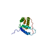
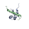
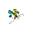
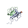
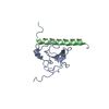
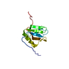
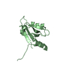
Mass: 7705.826 Da / Num. of mol.: 2 / Mutation: N11L, L12N Source method: isolated from a genetically manipulated source Source: (gene. exp.) Enterobacteria phage P22 (virus) / Genus: P22-like viruses / Gene: ARC / Plasmid: pET800 / Species (production host): Escherichia coli / Production host: Escherichia coli BL21(DE3) (bacteria) / Strain (production host): BL21(DE3) / References: UniProt: P03050
-
Experimental details
-
Experiment
Experiment
Method: SOLUTION NMR
NMR experiment
Conditions-ID
Experiment-ID
Solution-ID
Type
1
1
1
2D NOESY
1
2
1
3D 15N-separated NOESY
1
3
1
HNHA
1
4
1
HNHB
3
5
3
HSQCs (hydrogenexchange)
-
Sample preparation
Details
Solution-ID
Contents
Solvent system
1
4 mM uniform 15N-labelled Arc repressor NL11/LN12 (Switch Arc)
90% H2O/10% D2O
2
7.5 mM 10% 13C-labelled Arc repressor NL11/LN12 (Switch Arc)
90% H2O, 10% D20
3
5 mM uniform 15N-labelled Arc repressor NL11/LN12 (Switch Arc)
100% D2O
Sample conditions
Conditions-ID
Ionic strength
pH
Pressure (kPa)
Temperature (K)
1
20mMphosphate
4.8
ambient
303K
2
20mMphosphate
4.8
ambient
303K
3
20mMphosphate, 150mMKCl
4.67
ambient
303K
Crystal grow
*PLUS
Method: other / Details: NMR
-
NMR measurement
Radiation
Protocol: SINGLE WAVELENGTH / Monochromatic (M) / Laue (L): M
Radiation wavelength
Relative weight: 1
NMR spectrometer
Type: Bruker DMX / Manufacturer: Bruker / Model: DMX / Field strength: 500 MHz
-
Processing
NMR software
Name
Version
Developer
Classification
XwinNMR
2.1
Bruker
collection
NMRPipe
1.1
Delaglio, Grzesiek, Vuister, Zhu, Pfeifer, Bax
processing
NMRDraw
2.1
Delaglio, Grzesiek, Vuister, Zhu, Pfeifer, Bax
processing
NMRView
3.1
Johnston, Blevins
dataanalysis
X-PLOR
3.1
Brunger
structuresolution
X-PLOR
3.1
Brunger
refinement
Refinement
Method: simulated annealing / Software ordinal: 1 Details: Experimentally derived restraints included 810 nOe distance restraints per monomer, along with 3 hydrogen bond restraints, 31 phi-angle restraints and 3 chi1-angle restraints, for a total of ...Details: Experimentally derived restraints included 810 nOe distance restraints per monomer, along with 3 hydrogen bond restraints, 31 phi-angle restraints and 3 chi1-angle restraints, for a total of 847 restraints per monomer spanning residues 5-53. Structure calculations were performed using a simulated annealing protocol designed for symmetric dimers (M. Nilges, Proteins Struct. Funct. Gen, 17, 297 (1993)). As starting points for the calculation, 28 structures were generated in which the conformation of the N-terminal region (residues 1-13) was random, and that of the remainder of the protein restrained to be similar to wild-type Arc. The use of completely random starting structures led to extremely poor convergence rates, but those calculations which did converge yielded structures similar to those obtained from calculations using non-random starting structures. Each semi-random starting structure was then annealed to a model structure using the distance, angle and hydrogen bond restraints mentioned above. Two "seed" restraints, Arg 40 HE-Phe 45 HD and Trp 14 HE3-Tyr 38 HE, were described as unambiguously intermolecular. In the wild-type Arc structure, the difference between the intra and intermolecular distances for these atoms is >10 A. Use of the seed restraints improved convergence but again did not alter the qualitative nature of the results. All other NOE distance restraints were described ambiguously using sum potentials. Hydrogen-bond restraints in alpha-helices were described as intramonomer. In the conformational search phase of the calculations, non-bonded interactions were computed only between C-alpha atoms with a van der Waals term of 0.1. 13 of 28 calculated structures were accepted with no angle violations >5 degrees and no more than two nOe violations >0.35 A. These comprise the ensemble submitted here.
NMR representative
Selection criteria: closest to the average
NMR ensemble
Conformer selection criteria: structures with the least restraint violations Conformers calculated total number: 28 / Conformers submitted total number: 13
+
About Yorodumi
-
News
-
Feb 9, 2022. New format data for meta-information of EMDB entries
New format data for meta-information of EMDB entries
Version 3 of the EMDB header file is now the official format.
The previous official version 1.9 will be removed from the archive.
In the structure databanks used in Yorodumi, some data are registered as the other names, "COVID-19 virus" and "2019-nCoV". Here are the details of the virus and the list of structure data.
Jan 31, 2019. EMDB accession codes are about to change! (news from PDBe EMDB page)
EMDB accession codes are about to change! (news from PDBe EMDB page)
The allocation of 4 digits for EMDB accession codes will soon come to an end. Whilst these codes will remain in use, new EMDB accession codes will include an additional digit and will expand incrementally as the available range of codes is exhausted. The current 4-digit format prefixed with “EMD-” (i.e. EMD-XXXX) will advance to a 5-digit format (i.e. EMD-XXXXX), and so on. It is currently estimated that the 4-digit codes will be depleted around Spring 2019, at which point the 5-digit format will come into force.
The EM Navigator/Yorodumi systems omit the EMD- prefix.
Related info.:Q: What is EMD? / ID/Accession-code notation in Yorodumi/EM Navigator
Yorodumi is a browser for structure data from EMDB, PDB, SASBDB, etc.
This page is also the successor to EM Navigator detail page, and also detail information page/front-end page for Omokage search.
The word "yorodu" (or yorozu) is an old Japanese word meaning "ten thousand". "mi" (miru) is to see.
Related info.:EMDB / PDB / SASBDB / Comparison of 3 databanks / Yorodumi Search / Aug 31, 2016. New EM Navigator & Yorodumi / Yorodumi Papers / Jmol/JSmol / Function and homology information / Changes in new EM Navigator and Yorodumi
 Movie
Movie Controller

 Yorodumi
 Open data
 Basic information
 Components
 Keywords
 Function and homology information
 Enterobacteria phage P22 (virus)
 Authors
 Citation
 Structure visualization
 Downloads & links
 Other downloads
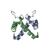

 PDBj
PDBj Assembly
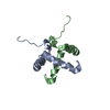

 Sample preparation
 Processing
 NMRPipe