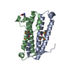
 Movie
Movie Controller

[English] 日本語
 Yorodumi
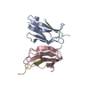
- PDB-1ng6: Structure of Cytosolic Protein of Unknown Function YqeY from Baci... -
+ Open data

- Basic information
| Entry | Database: PDB / ID: 1ng6 | ||||||
|---|---|---|---|---|---|---|---|
| Title | Structure of Cytosolic Protein of Unknown Function YqeY from Bacillus subtilis | ||||||
 Components Components | Hypothetical protein yqeY | ||||||
 Keywords Keywords | STRUCTURAL GENOMICS / UNKNOWN FUNCTION / Domain GatB/Yqey / pfam02637 / DUF186 / PSI / Protein Structure Initiative / Midwest Center for Structural Genomics / MCSG | ||||||
| Function / homology |  Function and homology information Function and homology information | ||||||
| Biological species |  | ||||||
| Method |  X-RAY DIFFRACTION / SYNCHROTRON / MAD / Resolution: 1.4 Å X-RAY DIFFRACTION / SYNCHROTRON / MAD / Resolution: 1.4 Å | ||||||
 Authors Authors | Zhang, R. / Dementiva, I. / Vinokour, E. / Collart, F. / Joachimiak, A. / Midwest Center for Structural Genomics (MCSG) | ||||||
 Citation Citation | Journal: To be Published Title: 1.4A crystal structure of hypothetical cytosolic protein YQEY Authors: Zhang, R. / Dementiva, I. / Vinokour, E. / Collart, F. / Joachimiak, A. | ||||||
| History |
|
- Structure visualization
| Structure viewer | Molecule: MolmilJmol/JSmol |
|---|
- Downloads & links
-Download
| PDBx/mmCIF format | 1ng6.cif.gz | 46.3 KB | Display | PDBx/mmCIF format |
|---|---|---|---|---|
| PDB format | pdb1ng6.ent.gz | 33 KB | Display | PDB format |
| PDBx/mmJSON format | 1ng6.json.gz | Tree view | PDBx/mmJSON format | |
| Others |  Other downloads Other downloads |
-Validation report
| Summary document | 1ng6_validation.pdf.gz | 351.5 KB | Display | wwPDB validaton report |
|---|---|---|---|---|
| Full document | 1ng6_full_validation.pdf.gz | 353.3 KB | Display | |
| Data in XML | 1ng6_validation.xml.gz | 4 KB | Display | |
| Data in CIF | 1ng6_validation.cif.gz | 7.3 KB | Display | |
| Arichive directory | https://data.pdbj.org/pub/pdb/validation_reports/ng/1ng6ftp://data.pdbj.org/pub/pdb/validation_reports/ng/1ng6 | HTTPS FTP |
-Related structure data
| Similar structure data | |
|---|---|
| Other databases |


-Links
 PDBj
PDBj- Assembly
| Deposited unit | 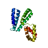
| ||||||||
|---|---|---|---|---|---|---|---|---|---|
| 1 |
| ||||||||
| Unit cell |
| ||||||||
| Details | This protein (APC1524) existed as monomer. One molecule in asymmetric unit. |
-Components
| #1: Protein | Mass: 16791.455 Da / Num. of mol.: 1 Source method: isolated from a genetically manipulated source Source: (gene. exp.) |
|---|---|
| #2: Water | ChemComp-HOH /  Mass: 18.015 Da / Num. of mol.: 295 / Source method: isolated from a natural source / Formula: H2O Mass: 18.015 Da / Num. of mol.: 295 / Source method: isolated from a natural source / Formula: H2O |
-Experimental details
-Experiment
| Experiment | Method: X-RAY DIFFRACTION / Number of used crystals: 1 |
|---|
- Sample preparation
| Crystal | Density Matthews: 2.1 Å3/Da / Density % sol: 41.44 % |
|---|---|
| Crystal grow | Temperature: 298 K / Method: vapor diffusion, hanging drop / pH: 5.5 Details: 19%PEG3350, 5% PEG400, 0.1M Bis Tris, pH 5.5, VAPOR DIFFUSION, HANGING DROP, temperature 298K |
-Data collection
| Diffraction | Mean temperature: 100 K | ||||||||||||
|---|---|---|---|---|---|---|---|---|---|---|---|---|---|
| Diffraction source | Source: SYNCHROTRON / Site: APS  / Beamline: 19-ID / Wavelength: 0.9795,0.9798,0.94656 / Beamline: 19-ID / Wavelength: 0.9795,0.9798,0.94656 | ||||||||||||
| Detector | Type: SBC-2 / Detector: CCD / Date: Dec 15, 2002 / Details: mirrors | ||||||||||||
| Radiation | Monochromator: Si 111 CHANNEL / Protocol: MAD / Monochromatic (M) / Laue (L): M / Scattering type: x-ray | ||||||||||||
| Radiation wavelength |
| ||||||||||||
| Reflection | Resolution: 1.4→50 Å / Num. all: 27607 / Num. obs: 27414 / % possible obs: 99.3 % / Observed criterion σ(F): 2 / Observed criterion σ(I): 2 / Redundancy: 6.95 % / Biso Wilson estimate: 13.4 Å2 / Rmerge(I) obs: 0.088 / Net I/σ(I): 30 | ||||||||||||
| Reflection shell | Resolution: 1.4→1.45 Å / Redundancy: 4.44 % / Rmerge(I) obs: 0.428 / Mean I/σ(I) obs: 2.4 / Num. unique all: 2661 / % possible all: 95.7 |
- Processing
| Software |
| ||||||||||||||||||||||||||||||||||||
|---|---|---|---|---|---|---|---|---|---|---|---|---|---|---|---|---|---|---|---|---|---|---|---|---|---|---|---|---|---|---|---|---|---|---|---|---|---|
| Refinement | Method to determine structure: MAD / Resolution: 1.4→34.71 Å / Rfactor Rfree error: 0.005 / Isotropic thermal model: RESTRAINED / Cross valid method: THROUGHOUT / σ(F): 0 / Stereochemistry target values: Engh & Huber Details: The number of reflections for refinement is greater than the number of reflections for data collection because in cns refinement (hlml target), the Fridel's pair was treated as two seperated reflections.
| ||||||||||||||||||||||||||||||||||||
| Solvent computation | Solvent model: FLAT MODEL / Bsol: 52.5572 Å2 / ksol: 0.336155 e/Å3 | ||||||||||||||||||||||||||||||||||||
| Displacement parameters | Biso mean: 17.6 Å2
| ||||||||||||||||||||||||||||||||||||
| Refine analyze | Luzzati coordinate error free: 0.2 Å / Luzzati sigma a free: 0.18 Å | ||||||||||||||||||||||||||||||||||||
| Refinement step | Cycle: LAST / Resolution: 1.4→34.71 Å
| ||||||||||||||||||||||||||||||||||||
| Refine LS restraints |
| ||||||||||||||||||||||||||||||||||||
| LS refinement shell | Resolution: 1.4→1.49 Å / Rfactor Rfree error: 0.016 / Total num. of bins used: 6
| ||||||||||||||||||||||||||||||||||||
| Xplor file |
|
