 Movie
Movie Controller


+ Open data

- Basic information
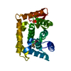
| Entry | Database: PDB / ID: 1ng5 | ||||||
|---|---|---|---|---|---|---|---|
| Title | 2.0 A crystal structure of Staphylococcus aureus Sortase B | ||||||
 Components Components | sortase B | ||||||
 Keywords Keywords | HYDROLASE / TRANSFERASE / structural genomics / a new fold / PSI / Protein Structure Initiative / Midwest Center for Structural Genomics / MCSG | ||||||
| Function / homology |  Function and homology information Function and homology information | ||||||
| Biological species |   Staphylococcus aureus subsp. aureus (bacteria) Staphylococcus aureus subsp. aureus (bacteria) | ||||||
| Method |  X-RAY DIFFRACTION / SYNCHROTRON / MAD / Resolution: 2 Å X-RAY DIFFRACTION / SYNCHROTRON / MAD / Resolution: 2 Å | ||||||
 Authors Authors | Zhang, R. / Joachimiak, G. / Joachimiak, A. / Midwest Center for Structural Genomics (MCSG) | ||||||
 Citation Citation | Journal: Structure / Year: 2004 Title: Structures of sortase B from Staphylococcus aureus and Bacillus anthracis reveal catalytic amino acid triad in the active site. Authors: Zhang, R. / Wu, R. / Joachimiak, G. / Mazmanian, S.K. / Missiakas, D.M. / Gornicki, P. / Schneewind, O. / Joachimiak, A. | ||||||
| History |
|
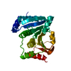
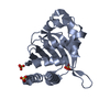
- Structure visualization
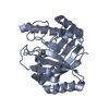
| Structure viewer | Molecule: MolmilJmol/JSmol |
|---|
- Downloads & links
-Download
| PDBx/mmCIF format | 1ng5.cif.gz | 103.3 KB | Display | PDBx/mmCIF format |
|---|---|---|---|---|
| PDB format | pdb1ng5.ent.gz | 80 KB | Display | PDB format |
| PDBx/mmJSON format | 1ng5.json.gz | Tree view | PDBx/mmJSON format | |
| Others |  Other downloads Other downloads |
-Validation report
| Arichive directory | https://data.pdbj.org/pub/pdb/validation_reports/ng/1ng5ftp://data.pdbj.org/pub/pdb/validation_reports/ng/1ng5 | HTTPS FTP |
|---|
-Related structure data


-Links
 PDBj
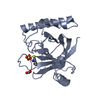
PDBj- Assembly
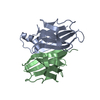
| Deposited unit | 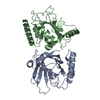
| ||||||||
|---|---|---|---|---|---|---|---|---|---|
| 1 | 
| ||||||||
| 2 | 
| ||||||||
| Unit cell |
| ||||||||
| Details | Sortase B existed as monomer, There are two molecules (A,B) in asymmetric unit. |
-Components
| #1: Protein | Mass: 25641.000 Da / Num. of mol.: 2 Source method: isolated from a genetically manipulated source Source: (gene. exp.) Staphylococcus aureus subsp. aureus (bacteria)Species: Staphylococcus aureus / Strain: N315 / Gene: SA0982 / Plasmid: PDM 68 / Species (production host): Escherichia coli / Production host: #2: Water | ChemComp-HOH / |  Mass: 18.015 Da / Num. of mol.: 307 / Source method: isolated from a natural source / Formula: H2O Mass: 18.015 Da / Num. of mol.: 307 / Source method: isolated from a natural source / Formula: H2O |
|---|
-Experimental details
-Experiment
| Experiment | Method: X-RAY DIFFRACTION / Number of used crystals: 2 |
|---|
- Sample preparation
| Crystal | Density Matthews: 2.11 Å3/Da / Density % sol: 41.59 % | |||||||||||||||||||||||||||||||||||||||||||||||||
|---|---|---|---|---|---|---|---|---|---|---|---|---|---|---|---|---|---|---|---|---|---|---|---|---|---|---|---|---|---|---|---|---|---|---|---|---|---|---|---|---|---|---|---|---|---|---|---|---|---|---|
| Crystal grow | Temperature: 298 K / Method: vapor diffusion, hanging drop / pH: 5.6 Details: 2.5M Na2SO4, 0.1M triNa citrate, pH 5.6, VAPOR DIFFUSION, HANGING DROP, temperature 298K | |||||||||||||||||||||||||||||||||||||||||||||||||
| Crystal grow | *PLUS pH: 7.5 / Method: vapor diffusion, hanging drop | |||||||||||||||||||||||||||||||||||||||||||||||||
| Components of the solutions | *PLUS
|
-Data collection
| Diffraction | Mean temperature: 100 K | ||||||||||||
|---|---|---|---|---|---|---|---|---|---|---|---|---|---|
| Diffraction source | Source: SYNCHROTRON / Site: APS  / Beamline: 19-ID / Wavelength: 0.9795,0.9798,0.94656 / Beamline: 19-ID / Wavelength: 0.9795,0.9798,0.94656 | ||||||||||||
| Detector | Type: SBC-2 / Detector: CCD / Date: Nov 19, 2001 / Details: mirrors | ||||||||||||
| Radiation | Monochromator: Si 111 channel / Protocol: MAD / Monochromatic (M) / Laue (L): M / Scattering type: x-ray | ||||||||||||
| Radiation wavelength |
| ||||||||||||
| Reflection | Resolution: 1.97→50 Å / Num. all: 31422 / Num. obs: 29674 / % possible obs: 94.4 % / Observed criterion σ(F): 4 / Observed criterion σ(I): 4 / Redundancy: 6 % / Biso Wilson estimate: 11.9 Å2 / Rmerge(I) obs: 0.11 / Net I/σ(I): 13 | ||||||||||||
| Reflection shell | Resolution: 1.97→2.05 Å / Redundancy: 4.81 % / Rmerge(I) obs: 0.537 / Mean I/σ(I) obs: 1.63 / Num. unique all: 2739 / % possible all: 79.5 | ||||||||||||
| Reflection | *PLUS Highest resolution: 2 Å / Rmerge(I) obs: 0.11 |
- Processing
| Software |
| |||||||||||||||||||||||||
|---|---|---|---|---|---|---|---|---|---|---|---|---|---|---|---|---|---|---|---|---|---|---|---|---|---|---|
| Refinement | Method to determine structure: MAD / Resolution: 2→42.09 Å / Rfactor Rfree error: 0.005 / Isotropic thermal model: RESTRAINED / Cross valid method: THROUGHOUT / σ(F): 0 / Stereochemistry target values: Engh & Huber Details: The number of reflections for refinement is greater than the number of reflections for data collection, because in CNS (hlml taget) refinement, the Friedel's pair was treated as two seperated reflections.
| |||||||||||||||||||||||||
| Solvent computation | Solvent model: FLAT MODEL / Bsol: 82.368 Å2 / ksol: 0.431764 e/Å3 | |||||||||||||||||||||||||
| Displacement parameters | Biso mean: 31.7 Å2
| |||||||||||||||||||||||||
| Refine analyze | Luzzati coordinate error free: 0.31 Å / Luzzati sigma a free: 0.39 Å | |||||||||||||||||||||||||
| Refinement step | Cycle: LAST / Resolution: 2→42.09 Å
| |||||||||||||||||||||||||
| Refine LS restraints |
| |||||||||||||||||||||||||
| LS refinement shell | Resolution: 2→2.13 Å / Rfactor Rfree error: 0.019 / Total num. of bins used: 6
| |||||||||||||||||||||||||
| Xplor file |
| |||||||||||||||||||||||||
| Refinement | *PLUS Highest resolution: 2 Å / Lowest resolution: 50 Å | |||||||||||||||||||||||||
| Solvent computation | *PLUS | |||||||||||||||||||||||||
| Displacement parameters | *PLUS | |||||||||||||||||||||||||
| Refine LS restraints | *PLUS
|
