 Movie
Movie Controller

[English] 日本語
 Yorodumi
- PDB-1nbh: Structure of glycine N-methyltransferase complexed with S-adenosy... -
+ Open data

- Basic information
| Entry | Database: PDB / ID: 1nbh | ||||||
|---|---|---|---|---|---|---|---|
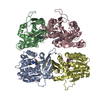
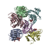
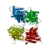
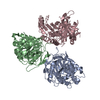
| Title | Structure of glycine N-methyltransferase complexed with S-adenosylmethionine and acetate, GNMT:SAM:Ace | ||||||
 Components Components | Glycine N-methyltransferase | ||||||
 Keywords Keywords | TRANSFERASE / methyltransferase / Glycine N-methyltransferase / S-adenosylmethionine / Enzyme catalytic mechanism | ||||||
| Function / homology |  Function and homology information Function and homology informationselenol Se-methyltransferase activity / Glyoxylate metabolism and glycine degradation / glycine N-methyltransferase / glycine N-methyltransferase activity / : / methyltransferase complex / L-methionine metabolic process / glycine metabolic process / : / S-adenosylmethionine metabolic process ...selenol Se-methyltransferase activity / Glyoxylate metabolism and glycine degradation / glycine N-methyltransferase / glycine N-methyltransferase activity / : / methyltransferase complex / L-methionine metabolic process / glycine metabolic process / : / S-adenosylmethionine metabolic process / S-adenosylmethionine-dependent methyltransferase activity / S-adenosyl-L-methionine binding / folic acid binding / regulation of gluconeogenesis / glycogen metabolic process / glycine binding / one-carbon metabolic process / methylation / protein homotetramerization / identical protein binding / cytosol Similarity search - Function | ||||||
| Biological species |  | ||||||
| Method |  X-RAY DIFFRACTION / MOLECULAR REPLACEMENT / Resolution: 2.8 Å X-RAY DIFFRACTION / MOLECULAR REPLACEMENT / Resolution: 2.8 Å | ||||||
 Authors Authors | Takata, Y. / Takusagawa, F. | ||||||
 Citation Citation | Journal: Biochemistry / Year: 2003 Title: Catalytic mechanism of glycine N-methyltransferase Authors: Takata, Y. / Huang, Y. / Komoto, J. / Yamada, T. / Konishi, K. / Ogawa, H. / Gomi, T. / Fujioka, M. / Takusagawa, F. | ||||||
| History |
|
- Structure visualization
| Structure viewer | Molecule: MolmilJmol/JSmol |
|---|
- Downloads & links
-Download
| PDBx/mmCIF format | 1nbh.cif.gz | 241.5 KB | Display | PDBx/mmCIF format |
|---|---|---|---|---|
| PDB format | pdb1nbh.ent.gz | 195.9 KB | Display | PDB format |
| PDBx/mmJSON format | 1nbh.json.gz | Tree view | PDBx/mmJSON format | |
| Others |  Other downloads Other downloads |
-Validation report
| Arichive directory | https://data.pdbj.org/pub/pdb/validation_reports/nb/1nbhftp://data.pdbj.org/pub/pdb/validation_reports/nb/1nbh | HTTPS FTP |
|---|
-Related structure data

-Links
 PDBj
PDBj
- Assembly
| Deposited unit | 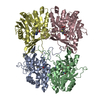
| ||||||||
|---|---|---|---|---|---|---|---|---|---|
| 1 |
| ||||||||
| Unit cell |
|
-Components
| #1: Protein | Mass: 32460.830 Da / Num. of mol.: 4 Source method: isolated from a genetically manipulated source Source: (gene. exp.)  #2: Chemical | ChemComp-ACT /   Mass: 59.044 Da / Num. of mol.: 4 / Source method: obtained synthetically / Formula: C2H3O2 Mass: 59.044 Da / Num. of mol.: 4 / Source method: obtained synthetically / Formula: C2H3O2#3: Chemical | ChemComp-SAM /   Mass: 398.437 Da / Num. of mol.: 4 / Source method: obtained synthetically / Formula: C15H22N6O5S Mass: 398.437 Da / Num. of mol.: 4 / Source method: obtained synthetically / Formula: C15H22N6O5S#4: Water | ChemComp-HOH / |  Mass: 18.015 Da / Num. of mol.: 289 / Source method: isolated from a natural source / Formula: H2O Mass: 18.015 Da / Num. of mol.: 289 / Source method: isolated from a natural source / Formula: H2O |
|---|
-Experimental details
-Experiment
| Experiment | Method: X-RAY DIFFRACTION / Number of used crystals: 1 |
|---|
- Sample preparation
| Crystal | Density Matthews: 2.79 Å3/Da / Density % sol: 55.98 % | |||||||||||||||||||||||||||||||||||||||||||||||||
|---|---|---|---|---|---|---|---|---|---|---|---|---|---|---|---|---|---|---|---|---|---|---|---|---|---|---|---|---|---|---|---|---|---|---|---|---|---|---|---|---|---|---|---|---|---|---|---|---|---|---|
| Crystal grow | Temperature: 296 K / Method: vapor diffusion, hanging drop / pH: 5.6 Details: PEG 3400, pH 5.6, VAPOR DIFFUSION, HANGING DROP, temperature 296K | |||||||||||||||||||||||||||||||||||||||||||||||||
| Crystal grow | *PLUS Temperature: 23 ℃ | |||||||||||||||||||||||||||||||||||||||||||||||||
| Components of the solutions | *PLUS
|
-Data collection
| Diffraction | Mean temperature: 100 K |
|---|---|
| Diffraction source | Source: ROTATING ANODE / Type: RIGAKU RU200 / Wavelength: 1.5418 Å |
| Detector | Type: RIGAKU RAXIS IIC / Detector: IMAGE PLATE / Date: Jan 10, 2002 / Details: Confocal optics |
| Radiation | Protocol: SINGLE WAVELENGTH / Monochromatic (M) / Laue (L): M / Scattering type: x-ray |
| Radiation wavelength | Wavelength: 1.5418 Å / Relative weight: 1 |
| Reflection | Resolution: 2.8→10 Å / Num. obs: 37008 / % possible obs: 90 % / Observed criterion σ(F): 0 / Observed criterion σ(I): 0 / Redundancy: 4.5 % / Rmerge(I) obs: 0.072 / Rsym value: 0.072 / Net I/σ(I): 5 |
| Reflection shell | Resolution: 2.8→3 Å / % possible all: 81 |
| Reflection | *PLUS Lowest resolution: 10 Å / Num. obs: 36870 / % possible obs: 99 % / Num. measured all: 195411 |
| Reflection shell | *PLUS Lowest resolution: 2.9 Å / % possible obs: 97 % / Rmerge(I) obs: 0.258 |
- Processing
| Software |
| ||||||||||||||||||||
|---|---|---|---|---|---|---|---|---|---|---|---|---|---|---|---|---|---|---|---|---|---|
| Refinement | Method to determine structure: MOLECULAR REPLACEMENT / Resolution: 2.8→10 Å / σ(F): 3 / Stereochemistry target values: Engh & Huber
| ||||||||||||||||||||
| Refinement step | Cycle: LAST / Resolution: 2.8→10 Å
| ||||||||||||||||||||
| Refine LS restraints |
| ||||||||||||||||||||
| Refinement | *PLUS Lowest resolution: 10 Å | ||||||||||||||||||||
| Solvent computation | *PLUS | ||||||||||||||||||||
| Displacement parameters | *PLUS | ||||||||||||||||||||
| Refine LS restraints | *PLUS
|
