 Movie
Movie Controller


+ Open data

- Basic information
| Entry | Database: PDB / ID: 1naq | ||||||
|---|---|---|---|---|---|---|---|
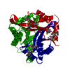
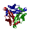
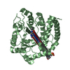
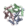
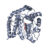
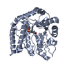
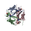
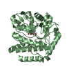
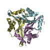
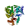
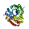
| Title | Crystal structure of CUTA1 from E.coli at 1.7 A resolution | ||||||
 Components Components | Periplasmic divalent cation tolerance protein cutA | ||||||
 Keywords Keywords | ELECTRON TRANSPORT / CUTA / copper resistance / Structural Proteomics in Europe / SPINE / Structural Genomics | ||||||
| Function / homology |  Function and homology information Function and homology informationresponse to copper ion / copper ion binding / protein-containing complex / metal ion binding / cytoplasm Similarity search - Function | ||||||
| Biological species |  | ||||||
| Method |  X-RAY DIFFRACTION / SYNCHROTRON / MAD / Resolution: 1.7 Å X-RAY DIFFRACTION / SYNCHROTRON / MAD / Resolution: 1.7 Å | ||||||
 Authors Authors | Calderone, V. / Mangani, S. / Benvenuti, M. / Viezzoli, M.S. / Banci, L. / Bertini, I. / Structural Proteomics in Europe (SPINE) | ||||||
 Citation Citation | Journal: J.Biol.Chem. / Year: 2003 Title: The evolutionarily conserved trimeric structure of CutA1 proteins suggests a role in signal transduction. Authors: Arnesano, F. / Banci, L. / Benvenuti, M. / Bertini, I. / Calderone, V. / Mangani, S. / Viezzoli, M.S. #1: Journal: To be PublishedTitle: STRUCTURE OF PROTEIN TM1056, CUTA Authors: SAVCHENKO, A. / ZHANG, R. / JOACHIMIAK, A. / EDWARDS, A. / AKARINA, T. | ||||||
| History |
| ||||||
| Remark 600 | HETEROGEN P-HYDROXYMERCURIBENZOIC ACID HAS BEEN ADDED TO THE PROTEIN PRIOR TO CRYSTALLISATION. IT ...HETEROGEN P-HYDROXYMERCURIBENZOIC ACID HAS BEEN ADDED TO THE PROTEIN PRIOR TO CRYSTALLISATION. IT REACTS WITH THE -SH OF FREE CYSTEINS AND, BY THE ELIMINATION OF ONE WATER MOLECULE, FORMS A COVALENT BOND BETWEEN THE S OF THE CYS AND HG WHICH IS THEN A GOOD CANDIDATE TO PERFORM A MAD EXPERIMENT. |
- Structure visualization
| Structure viewer | Molecule: MolmilJmol/JSmol |
|---|
- Downloads & links
-Download
| PDBx/mmCIF format | 1naq.cif.gz | 143.5 KB | Display | PDBx/mmCIF format |
|---|---|---|---|---|
| PDB format | pdb1naq.ent.gz | 114.3 KB | Display | PDB format |
| PDBx/mmJSON format | 1naq.json.gz | Tree view | PDBx/mmJSON format | |
| Others |  Other downloads Other downloads |
-Validation report
| Summary document | 1naq_validation.pdf.gz | 2.6 MB | Display | wwPDB validaton report |
|---|---|---|---|---|
| Full document | 1naq_full_validation.pdf.gz | 2.5 MB | Display | |
| Data in XML | 1naq_validation.xml.gz | 34.6 KB | Display | |
| Data in CIF | 1naq_validation.cif.gz | 46 KB | Display | |
| Arichive directory | https://data.pdbj.org/pub/pdb/validation_reports/na/1naqftp://data.pdbj.org/pub/pdb/validation_reports/na/1naq | HTTPS FTP |
-Related structure data

-Links
 PDBj
PDBj


- Assembly
| Deposited unit | 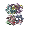
| ||||||||
|---|---|---|---|---|---|---|---|---|---|
| 1 | 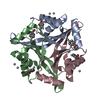
| ||||||||
| 2 | 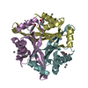
| ||||||||
| 3 |
| ||||||||
| Unit cell |
| ||||||||
| Details | The biological functional unit is a trimer; the asymmetric unit is made of two trimers. |
-Components
| #1: Protein | Mass: 12342.025 Da / Num. of mol.: 6 Source method: isolated from a genetically manipulated source Source: (gene. exp.) #2: Chemical | ChemComp-HG /   Mass: 200.590 Da / Num. of mol.: 8 / Source method: obtained synthetically / Formula: Hg Mass: 200.590 Da / Num. of mol.: 8 / Source method: obtained synthetically / Formula: Hg#3: Chemical | ChemComp-MBO /   Mass: 321.703 Da / Num. of mol.: 10 / Source method: obtained synthetically / Formula: C7H5HgO2 Mass: 321.703 Da / Num. of mol.: 10 / Source method: obtained synthetically / Formula: C7H5HgO2#4: Water | ChemComp-HOH / |  Mass: 18.015 Da / Num. of mol.: 343 / Source method: isolated from a natural source / Formula: H2O Mass: 18.015 Da / Num. of mol.: 343 / Source method: isolated from a natural source / Formula: H2O |
|---|
-Experimental details
-Experiment
| Experiment | Method: X-RAY DIFFRACTION / Number of used crystals: 1 |
|---|
- Sample preparation
| Crystal | Density Matthews: 2.07 Å3/Da / Density % sol: 40.56 % | ||||||||||||||||||||||||||||||||||||
|---|---|---|---|---|---|---|---|---|---|---|---|---|---|---|---|---|---|---|---|---|---|---|---|---|---|---|---|---|---|---|---|---|---|---|---|---|---|
| Crystal grow | Temperature: 293 K / Method: vapor diffusion, sitting drop / pH: 7.5 Details: 0.1M Na HEPES, 2M Ammonium sulphate, 2% PEG 400, 2 mM 4-(hydroxymercuri)benzoic acid, pH 7.5, VAPOR DIFFUSION, SITTING DROP, temperature 293K | ||||||||||||||||||||||||||||||||||||
| Crystal grow | *PLUS Temperature: 20 ℃ / Method: vapor diffusion | ||||||||||||||||||||||||||||||||||||
| Components of the solutions | *PLUS
|
-Data collection
| Diffraction | Mean temperature: 100 K | ||||||||||||
|---|---|---|---|---|---|---|---|---|---|---|---|---|---|
| Diffraction source | Source: SYNCHROTRON / Site: EMBL/DESY, HAMBURG  / Beamline: BW7A / Wavelength: 1.005231, 1.00870, 0.93200 / Beamline: BW7A / Wavelength: 1.005231, 1.00870, 0.93200 | ||||||||||||
| Detector | Type: MARRESEARCH / Detector: CCD / Date: Aug 14, 2002 | ||||||||||||
| Radiation | Monochromator: Double crystal focussing monochromator / Protocol: MAD / Monochromatic (M) / Laue (L): M / Scattering type: x-ray | ||||||||||||
| Radiation wavelength |
| ||||||||||||
| Reflection | Resolution: 1.7→37.5 Å / Num. all: 65739 / Num. obs: 65739 / % possible obs: 97.2 % / Observed criterion σ(F): 0 / Observed criterion σ(I): 0 / Redundancy: 9.7 % / Biso Wilson estimate: 18.95 Å2 / Rmerge(I) obs: 0.091 / Rsym value: 0.091 / Net I/σ(I): 5.7 | ||||||||||||
| Reflection shell | Resolution: 1.7→1.79 Å / Redundancy: 9.6 % / Rmerge(I) obs: 0.619 / Mean I/σ(I) obs: 1.1 / Num. unique all: 9337 / Rsym value: 0.619 / % possible all: 96.3 | ||||||||||||
| Reflection | *PLUS Lowest resolution: 40 Å / Num. measured all: 643844 | ||||||||||||
| Reflection shell | *PLUS % possible obs: 96.3 % / Num. unique obs: 9337 / Num. measured obs: 90452 / Rmerge(I) obs: 0.519 |
- Processing
| Software |
| ||||||||||||||||||||||||||||||||||||||||||||||||||||||||||||
|---|---|---|---|---|---|---|---|---|---|---|---|---|---|---|---|---|---|---|---|---|---|---|---|---|---|---|---|---|---|---|---|---|---|---|---|---|---|---|---|---|---|---|---|---|---|---|---|---|---|---|---|---|---|---|---|---|---|---|---|---|---|
| Refinement | Method to determine structure: MAD / Resolution: 1.7→19.96 Å / Cor.coef. Fo:Fc: 0.938 / Cor.coef. Fo:Fc free: 0.903 / SU B: 3.338 / SU ML: 0.11 / Cross valid method: THROUGHOUT / σ(F): 0 / σ(I): 0 / ESU R: 0.164 / ESU R Free: 0.152 / Stereochemistry target values: MAXIMUM LIKELIHOOD Details: At the N-terminus of all the six molecules present in the asymetric unit there are about 6-8 residues for which it's not possible to see a clear density. Among the densities belonging to ...Details: At the N-terminus of all the six molecules present in the asymetric unit there are about 6-8 residues for which it's not possible to see a clear density. Among the densities belonging to each asymmetric unit it's possible to see some extra density which could account for the presence of some crystalline PEG fragments.
| ||||||||||||||||||||||||||||||||||||||||||||||||||||||||||||
| Solvent computation | Ion probe radii: 0.8 Å / Shrinkage radii: 0.8 Å / VDW probe radii: 1.4 Å / Solvent model: BABINET MODEL WITH MASK | ||||||||||||||||||||||||||||||||||||||||||||||||||||||||||||
| Displacement parameters | Biso mean: 20.738 Å2
| ||||||||||||||||||||||||||||||||||||||||||||||||||||||||||||
| Refine analyze | Luzzati sigma a obs: 0.055714 Å | ||||||||||||||||||||||||||||||||||||||||||||||||||||||||||||
| Refinement step | Cycle: LAST / Resolution: 1.7→19.96 Å
| ||||||||||||||||||||||||||||||||||||||||||||||||||||||||||||
| Refine LS restraints |
| ||||||||||||||||||||||||||||||||||||||||||||||||||||||||||||
| LS refinement shell | Resolution: 1.7→1.79 Å / Total num. of bins used: 20
| ||||||||||||||||||||||||||||||||||||||||||||||||||||||||||||
| Refinement | *PLUS Highest resolution: 1.7 Å / Lowest resolution: 20 Å / Rfactor Rfree: 0.255 / Rfactor Rwork: 0.203 | ||||||||||||||||||||||||||||||||||||||||||||||||||||||||||||
| Solvent computation | *PLUS | ||||||||||||||||||||||||||||||||||||||||||||||||||||||||||||
| Displacement parameters | *PLUS | ||||||||||||||||||||||||||||||||||||||||||||||||||||||||||||
| Refine LS restraints | *PLUS
|
