 Movie
Movie Controller

[English] 日本語
 Yorodumi
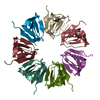
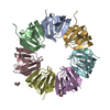
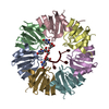
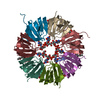
- PDB-1n9r: Crystal structure of a heptameric ring complex of yeast SmF in sp... -
+ Open data

- Basic information
| Entry | Database: PDB / ID: 1n9r | ||||||
|---|---|---|---|---|---|---|---|
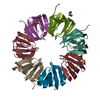
| Title | Crystal structure of a heptameric ring complex of yeast SmF in spacegroup P4122 | ||||||
 Components Components | Small nuclear ribonucleoprotein F | ||||||
 Keywords Keywords | TRANSLATION / snrnp / Sm protein / heptamer | ||||||
| Function / homology |  Function and homology information Function and homology informationU4/U6 snRNP / 7-methylguanosine cap hypermethylation / pICln-Sm protein complex / splicing factor binding / commitment complex / U2-type prespliceosome assembly / U1 snRNP / U2 snRNP / U4 snRNP / poly(U) RNA binding ...U4/U6 snRNP / 7-methylguanosine cap hypermethylation / pICln-Sm protein complex / splicing factor binding / commitment complex / U2-type prespliceosome assembly / U1 snRNP / U2 snRNP / U4 snRNP / poly(U) RNA binding / mRNA 5'-splice site recognition / spliceosomal complex assembly / Prp19 complex / U5 snRNP / spliceosomal snRNP assembly / U4/U6 x U5 tri-snRNP complex / catalytic step 2 spliceosome / spliceosomal complex / mRNA splicing, via spliceosome / RNA binding / nucleus / cytoplasm Similarity search - Function | ||||||
| Biological species |  | ||||||
| Method |  X-RAY DIFFRACTION / SYNCHROTRON / MOLECULAR REPLACEMENT / Resolution: 2.8 Å X-RAY DIFFRACTION / SYNCHROTRON / MOLECULAR REPLACEMENT / Resolution: 2.8 Å | ||||||
 Authors Authors | Collins, B.M. / Cubeddu, L. / Naidoo, N. / Harrop, S.J. / Kornfeld, G.D. / Dawes, I.W. / Curmi, P.M.G. / Mabbutt, B.C. | ||||||
 Citation Citation | Journal: J.Biol.Chem. / Year: 2003 Title: Homomeric ring assemblies of eukaryotic Sm proteins have affinity for both RNA and DNA: Crystal structure of an oligomeric complex of yeast SmF Authors: Collins, B.M. / Cubeddu, L. / Naidoo, N. / Harrop, S.J. / Kornfeld, G.D. / Dawes, I.W. / Curmi, P.M.G. / Mabbutt, B.C. | ||||||
| History |
|
- Structure visualization
| Structure viewer | Molecule: MolmilJmol/JSmol |
|---|
- Downloads & links
-Download
| PDBx/mmCIF format | 1n9r.cif.gz | 106 KB | Display | PDBx/mmCIF format |
|---|---|---|---|---|
| PDB format | pdb1n9r.ent.gz | 84.3 KB | Display | PDB format |
| PDBx/mmJSON format | 1n9r.json.gz | Tree view | PDBx/mmJSON format | |
| Others |  Other downloads Other downloads |
-Validation report
| Arichive directory | https://data.pdbj.org/pub/pdb/validation_reports/n9/1n9rftp://data.pdbj.org/pub/pdb/validation_reports/n9/1n9r | HTTPS FTP |
|---|
-Related structure data
| Related structure data | 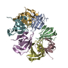 1n9sC 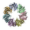 1i81S S: Starting model for refinement C: citing same article ( |
|---|---|
| Similar structure data |


-Links
 PDBj
PDBj

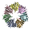
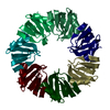
- Assembly
| Deposited unit | 
| ||||||||||
|---|---|---|---|---|---|---|---|---|---|---|---|
| 1 |
| ||||||||||
| 2 | 
| ||||||||||
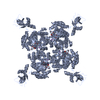
| Unit cell |
|
-Components
| #1: Protein | Mass: 10630.022 Da / Num. of mol.: 7 Source method: isolated from a genetically manipulated source Source: (gene. exp.) Production host:  |
|---|
-Experimental details
-Experiment
| Experiment | Method: X-RAY DIFFRACTION / Number of used crystals: 1 |
|---|
- Sample preparation
| Crystal | Density Matthews: 2.69 Å3/Da / Density % sol: 54.31 % | ||||||||||||||||||||||||||||||||||||||||||
|---|---|---|---|---|---|---|---|---|---|---|---|---|---|---|---|---|---|---|---|---|---|---|---|---|---|---|---|---|---|---|---|---|---|---|---|---|---|---|---|---|---|---|---|
| Crystal grow | Temperature: 289 K / Method: vapor diffusion, sitting drop / pH: 8.5 Details: PEG 3350, sodium acetate, pH 8.5, VAPOR DIFFUSION, SITTING DROP at 289K | ||||||||||||||||||||||||||||||||||||||||||
| Crystal grow | *PLUS | ||||||||||||||||||||||||||||||||||||||||||
| Components of the solutions | *PLUS
|
-Data collection
| Diffraction | Mean temperature: 100 K |
|---|---|
| Diffraction source | Source: SYNCHROTRON / Site: ESRF  / Beamline: ID14-4 / Wavelength: 0.9393 Å / Beamline: ID14-4 / Wavelength: 0.9393 Å |
| Detector | Detector: AREA DETECTOR |
| Radiation | Protocol: SINGLE WAVELENGTH / Monochromatic (M) / Laue (L): M / Scattering type: x-ray |
| Radiation wavelength | Wavelength: 0.9393 Å / Relative weight: 1 |
| Reflection | Resolution: 2.8→79 Å / Num. all: 19819 / % possible obs: 99.9 % / Redundancy: 6.8 % / Biso Wilson estimate: 84.2 Å2 / Rmerge(I) obs: 0.066 / Net I/σ(I): 19.6 |
| Reflection shell | Resolution: 2.8→2.95 Å / Redundancy: 7.1 % / Rmerge(I) obs: 0.39 / Mean I/σ(I) obs: 4.6 |
| Reflection | *PLUS Highest resolution: 2.8 Å |
| Reflection shell | *PLUS % possible obs: 99.9 % / Rmerge(I) obs: 0.39 |
- Processing
| Software |
| ||||||||||||||||||||||||||||||||||||||||||||||||||||||||||||||||||||||||||||||||||||||||||||||||||||||||||||||||||||||||||||||||||||||||||||||||||||||||||||||||||||||||||||||||||||||||||||||||||||||||
|---|---|---|---|---|---|---|---|---|---|---|---|---|---|---|---|---|---|---|---|---|---|---|---|---|---|---|---|---|---|---|---|---|---|---|---|---|---|---|---|---|---|---|---|---|---|---|---|---|---|---|---|---|---|---|---|---|---|---|---|---|---|---|---|---|---|---|---|---|---|---|---|---|---|---|---|---|---|---|---|---|---|---|---|---|---|---|---|---|---|---|---|---|---|---|---|---|---|---|---|---|---|---|---|---|---|---|---|---|---|---|---|---|---|---|---|---|---|---|---|---|---|---|---|---|---|---|---|---|---|---|---|---|---|---|---|---|---|---|---|---|---|---|---|---|---|---|---|---|---|---|---|---|---|---|---|---|---|---|---|---|---|---|---|---|---|---|---|---|---|---|---|---|---|---|---|---|---|---|---|---|---|---|---|---|---|---|---|---|---|---|---|---|---|---|---|---|---|---|---|---|---|
| Refinement | Method to determine structure: MOLECULAR REPLACEMENT Starting model: PDB entry 1I81 truncated to poly-serine Resolution: 2.8→20 Å / Cor.coef. Fo:Fc: 0.916 / Cor.coef. Fo:Fc free: 0.911 / TLS residual ADP flag: LIKELY RESIDUAL / Isotropic thermal model: TLS refinement used / Cross valid method: THROUGHOUT / σ(F): 0 / Stereochemistry target values: MAXIMUM LIKELIHOOD / Details: HYDROGENS HAVE BEEN ADDED IN THE RIDING POSITIONS
| ||||||||||||||||||||||||||||||||||||||||||||||||||||||||||||||||||||||||||||||||||||||||||||||||||||||||||||||||||||||||||||||||||||||||||||||||||||||||||||||||||||||||||||||||||||||||||||||||||||||||
| Solvent computation | Shrinkage radii: 0.8 Å / Solvent model: BABINET MODEL WITH MASK | ||||||||||||||||||||||||||||||||||||||||||||||||||||||||||||||||||||||||||||||||||||||||||||||||||||||||||||||||||||||||||||||||||||||||||||||||||||||||||||||||||||||||||||||||||||||||||||||||||||||||
| Displacement parameters | Biso mean: 50.316 Å2
| ||||||||||||||||||||||||||||||||||||||||||||||||||||||||||||||||||||||||||||||||||||||||||||||||||||||||||||||||||||||||||||||||||||||||||||||||||||||||||||||||||||||||||||||||||||||||||||||||||||||||
| Refinement step | Cycle: LAST / Resolution: 2.8→20 Å
| ||||||||||||||||||||||||||||||||||||||||||||||||||||||||||||||||||||||||||||||||||||||||||||||||||||||||||||||||||||||||||||||||||||||||||||||||||||||||||||||||||||||||||||||||||||||||||||||||||||||||
| Refine LS restraints |
| ||||||||||||||||||||||||||||||||||||||||||||||||||||||||||||||||||||||||||||||||||||||||||||||||||||||||||||||||||||||||||||||||||||||||||||||||||||||||||||||||||||||||||||||||||||||||||||||||||||||||
| LS refinement shell | Resolution: 2.8→2.871 Å / Total num. of bins used: 20 /
| ||||||||||||||||||||||||||||||||||||||||||||||||||||||||||||||||||||||||||||||||||||||||||||||||||||||||||||||||||||||||||||||||||||||||||||||||||||||||||||||||||||||||||||||||||||||||||||||||||||||||
| Refinement TLS params. | Method: refined / Refine-ID: X-RAY DIFFRACTION
| ||||||||||||||||||||||||||||||||||||||||||||||||||||||||||||||||||||||||||||||||||||||||||||||||||||||||||||||||||||||||||||||||||||||||||||||||||||||||||||||||||||||||||||||||||||||||||||||||||||||||
| Refinement TLS group |
| ||||||||||||||||||||||||||||||||||||||||||||||||||||||||||||||||||||||||||||||||||||||||||||||||||||||||||||||||||||||||||||||||||||||||||||||||||||||||||||||||||||||||||||||||||||||||||||||||||||||||
| Refinement | *PLUS Highest resolution: 2.8 Å / Lowest resolution: 20 Å / Rfactor Rfree: 0.267 / Rfactor Rwork: 0.254 | ||||||||||||||||||||||||||||||||||||||||||||||||||||||||||||||||||||||||||||||||||||||||||||||||||||||||||||||||||||||||||||||||||||||||||||||||||||||||||||||||||||||||||||||||||||||||||||||||||||||||
| Solvent computation | *PLUS | ||||||||||||||||||||||||||||||||||||||||||||||||||||||||||||||||||||||||||||||||||||||||||||||||||||||||||||||||||||||||||||||||||||||||||||||||||||||||||||||||||||||||||||||||||||||||||||||||||||||||
| Displacement parameters | *PLUS | ||||||||||||||||||||||||||||||||||||||||||||||||||||||||||||||||||||||||||||||||||||||||||||||||||||||||||||||||||||||||||||||||||||||||||||||||||||||||||||||||||||||||||||||||||||||||||||||||||||||||
| Refine LS restraints | *PLUS
|
