 Movie
Movie Controller

[English] 日本語
 Yorodumi
- PDB-1n1d: Glycerol-3-phosphate cytidylyltransferase complexed with CDP-glycerol -
+ Open data

- Basic information
| Entry | Database: PDB / ID: 1n1d | ||||||
|---|---|---|---|---|---|---|---|
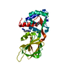
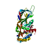
| Title | Glycerol-3-phosphate cytidylyltransferase complexed with CDP-glycerol | ||||||
 Components Components | glycerol-3-phosphate cytidylyltransferase | ||||||
 Keywords Keywords | TRANSFERASE / alpha/beta fold / cytidylyltransferase / nucleotidyltransferase / negative cooperativity / CDP-glycerol | ||||||
| Function / homology |  Function and homology information Function and homology informationglycerol-3-phosphate cytidylyltransferase / glycerol-3-phosphate cytidylyltransferase activity / teichoic acid biosynthetic process / cell wall organization / metal ion binding / cytoplasm Similarity search - Function | ||||||
| Biological species |  | ||||||
| Method |  X-RAY DIFFRACTION / MOLECULAR REPLACEMENT / Resolution: 2 Å X-RAY DIFFRACTION / MOLECULAR REPLACEMENT / Resolution: 2 Å | ||||||
 Authors Authors | Pattridge, K.A. / Weber, C.H. / Friesen, J.A. / Sankar, S. / Kent, C. / Ludwig, M.L. | ||||||
 Citation Citation | Journal: J.Biol.Chem. / Year: 2003 Title: Glycerol-3-phosphate cytidylyltransferase. Structural changes induced by binding of CDP-glycerol and the role of lysine residues in catalysis Authors: Pattridge, K.A. / Weber, C.H. / Friesen, J.A. / Sanker, S. / Kent, C. / Ludwig, M.L. #1: Journal: Structure / Year: 1999Title: A prototypical cytidylyltransferase: CTP:glycerol-3-phosphate cytidylyltransferase from Bacillus subtilis Authors: Weber, C.H. / Park, Y.S. / Sanker, S. / Kent, C. / Ludwig, M.L. | ||||||
| History |
|
- Structure visualization
| Structure viewer | Molecule: MolmilJmol/JSmol |
|---|
- Downloads & links
-Download
| PDBx/mmCIF format | 1n1d.cif.gz | 120.7 KB | Display | PDBx/mmCIF format |
|---|---|---|---|---|
| PDB format | pdb1n1d.ent.gz | 95.6 KB | Display | PDB format |
| PDBx/mmJSON format | 1n1d.json.gz | Tree view | PDBx/mmJSON format | |
| Others |  Other downloads Other downloads |
-Validation report
| Summary document | 1n1d_validation.pdf.gz | 1.5 MB | Display | wwPDB validaton report |
|---|---|---|---|---|
| Full document | 1n1d_full_validation.pdf.gz | 1.5 MB | Display | |
| Data in XML | 1n1d_validation.xml.gz | 24.4 KB | Display | |
| Data in CIF | 1n1d_validation.cif.gz | 32.2 KB | Display | |
| Arichive directory | https://data.pdbj.org/pub/pdb/validation_reports/n1/1n1dftp://data.pdbj.org/pub/pdb/validation_reports/n1/1n1d | HTTPS FTP |
-Related structure data
| Related structure data |  1cozS S: Starting model for refinement |
|---|---|
| Similar structure data |


-Links
 PDBj
PDBj- Assembly
| Deposited unit | 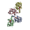
| ||||||||
|---|---|---|---|---|---|---|---|---|---|
| 1 | 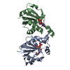
| ||||||||
| 2 | 
| ||||||||
| Unit cell |
|
-Components
| #1: Protein | Mass: 15295.539 Da / Num. of mol.: 4 Source method: isolated from a genetically manipulated source Source: (gene. exp.) References: UniProt: P27623, glycerol-3-phosphate cytidylyltransferase #2: Chemical |   Mass: 96.063 Da / Num. of mol.: 3 / Source method: obtained synthetically / Formula: SO4 Mass: 96.063 Da / Num. of mol.: 3 / Source method: obtained synthetically / Formula: SO4#3: Chemical | ChemComp-C2G / [   Mass: 477.255 Da / Num. of mol.: 4 / Source method: obtained synthetically / Formula: C12H21N3O13P2 Mass: 477.255 Da / Num. of mol.: 4 / Source method: obtained synthetically / Formula: C12H21N3O13P2#4: Water | ChemComp-HOH / |  Mass: 18.015 Da / Num. of mol.: 169 / Source method: isolated from a natural source / Formula: H2O Mass: 18.015 Da / Num. of mol.: 169 / Source method: isolated from a natural source / Formula: H2O |
|---|
-Experimental details
-Experiment
| Experiment | Method: X-RAY DIFFRACTION / Number of used crystals: 1 |
|---|
- Sample preparation
| Crystal | Density Matthews: 2.03 Å3/Da / Density % sol: 38.8 % | |||||||||||||||||||||||||||||||||||||||||||||||||||||||||||||||
|---|---|---|---|---|---|---|---|---|---|---|---|---|---|---|---|---|---|---|---|---|---|---|---|---|---|---|---|---|---|---|---|---|---|---|---|---|---|---|---|---|---|---|---|---|---|---|---|---|---|---|---|---|---|---|---|---|---|---|---|---|---|---|---|---|
| Crystal grow | Temperature: 298 K / Method: vapor diffusion, hanging drop / pH: 8.5 Details: PEG 3350, lithium sulfate, Tris, EDTA, DTT, pH 8.5, VAPOR DIFFUSION, HANGING DROP, temperature 298K | |||||||||||||||||||||||||||||||||||||||||||||||||||||||||||||||
| Crystal grow | *PLUS Method: vapor diffusion | |||||||||||||||||||||||||||||||||||||||||||||||||||||||||||||||
| Components of the solutions | *PLUS
|
-Data collection
| Diffraction | Mean temperature: 140 K |
|---|---|
| Diffraction source | Source: ROTATING ANODE / Type: RIGAKU / Wavelength: 1.5418 Å |
| Detector | Type: RIGAKU RAXIS IV / Detector: IMAGE PLATE / Date: Dec 10, 1998 / Details: mirrors |
| Radiation | Monochromator: YALE MIRRORS / Protocol: SINGLE WAVELENGTH / Monochromatic (M) / Laue (L): M / Scattering type: x-ray |
| Radiation wavelength | Wavelength: 1.5418 Å / Relative weight: 1 |
| Reflection | Resolution: 1.8→99 Å / Num. all: 40625 / Num. obs: 40625 / % possible obs: 87.8 % / Observed criterion σ(F): 0 / Observed criterion σ(I): 0 / Redundancy: 3 % / Rsym value: 0.051 / Net I/σ(I): 20.6 |
| Reflection shell | Resolution: 1.8→1.87 Å / Mean I/σ(I) obs: 5.7 / Rsym value: 0.191 / % possible all: 82.1 |
| Reflection | *PLUS Num. obs: 40610 / Num. measured all: 121437 / Rmerge(I) obs: 0.051 |
| Reflection shell | *PLUS Highest resolution: 1.8 Å / % possible obs: 82.1 % / Rmerge(I) obs: 0.191 |
- Processing
| Software |
| ||||||||||||||||||||||||||||||||||||
|---|---|---|---|---|---|---|---|---|---|---|---|---|---|---|---|---|---|---|---|---|---|---|---|---|---|---|---|---|---|---|---|---|---|---|---|---|---|
| Refinement | Method to determine structure: MOLECULAR REPLACEMENT Starting model: PDB ENTRY 1COZ Resolution: 2→15 Å / Cross valid method: THROUGHOUT / σ(F): 0 / σ(I): 0 / Stereochemistry target values: Engh & Huber
| ||||||||||||||||||||||||||||||||||||
| Displacement parameters | Biso mean: 12.1 Å2
| ||||||||||||||||||||||||||||||||||||
| Refine analyze |
| ||||||||||||||||||||||||||||||||||||
| Refinement step | Cycle: LAST / Resolution: 2→15 Å
| ||||||||||||||||||||||||||||||||||||
| Refine LS restraints |
| ||||||||||||||||||||||||||||||||||||
| Xplor file |
| ||||||||||||||||||||||||||||||||||||
| Refinement | *PLUS Highest resolution: 1.8 Å / Lowest resolution: 15 Å / Num. reflection obs: 40610 / Num. reflection Rfree: 2032 | ||||||||||||||||||||||||||||||||||||
| Solvent computation | *PLUS | ||||||||||||||||||||||||||||||||||||
| Displacement parameters | *PLUS | ||||||||||||||||||||||||||||||||||||
| Refine LS restraints | *PLUS
|
