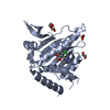
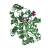
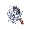
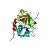
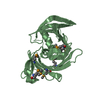
 Movie
Movie Controller


+ Open data

- Basic information
| Entry | Database: PDB / ID: 1mzd | ||||||
|---|---|---|---|---|---|---|---|
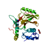
| Title | crystal structure of human pro-granzyme K | ||||||
 Components Components | pro-granzyme K | ||||||
 Keywords Keywords | HYDROLASE / granzyme / apoptosis / serine protease / S1 family | ||||||
| Function / homology |  Function and homology information Function and homology informationgranzyme-mediated programmed cell death signaling pathway / opsonization / complement activation, GZMK pathway / glycosaminoglycan binding / symbiont cell surface / zymogen activation / pattern recognition receptor activity / positive regulation of release of cytochrome c from mitochondria / Hydrolases; Acting on peptide bonds (peptidases); Serine endopeptidases / serine-type peptidase activity ...granzyme-mediated programmed cell death signaling pathway / opsonization / complement activation, GZMK pathway / glycosaminoglycan binding / symbiont cell surface / zymogen activation / pattern recognition receptor activity / positive regulation of release of cytochrome c from mitochondria / Hydrolases; Acting on peptide bonds (peptidases); Serine endopeptidases / serine-type peptidase activity / protein maturation / positive regulation of inflammatory response / serine-type endopeptidase activity / : Similarity search - Function | ||||||
| Biological species |  Homo sapiens (human) Homo sapiens (human) | ||||||
| Method |  X-RAY DIFFRACTION / FOURIER SYNTHESIS / Resolution: 2.9 Å X-RAY DIFFRACTION / FOURIER SYNTHESIS / Resolution: 2.9 Å | ||||||
 Authors Authors | Hink-Schauer, C. / Estebanez-Perpina, E. / Wilharm, E. / Fuentes-Prior, P. / Klinkert, W. / Bode, W. / Jenne, D.E. | ||||||
 Citation Citation | Journal: J.BIOL.CHEM. / Year: 2002 Title: The 2.2-A Crystal Structure of Human Pro-granzyme K Reveals a Rigid Zymogen with Unusual Features Authors: Hink-Schauer, C. / Estebanez-Perpina, E. / Wilharm, E. / Fuentes-Prior, P. / Klinkert, W. / Bode, W. / Jenne, D.E. | ||||||
| History |
|
- Structure visualization
| Structure viewer | Molecule: MolmilJmol/JSmol |
|---|
- Downloads & links
-Download
| PDBx/mmCIF format | 1mzd.cif.gz | 59.5 KB | Display | PDBx/mmCIF format |
|---|---|---|---|---|
| PDB format | pdb1mzd.ent.gz | 42.2 KB | Display | PDB format |
| PDBx/mmJSON format | 1mzd.json.gz | Tree view | PDBx/mmJSON format | |
| Others |  Other downloads Other downloads |
-Validation report
| Arichive directory | https://data.pdbj.org/pub/pdb/validation_reports/mz/1mzdftp://data.pdbj.org/pub/pdb/validation_reports/mz/1mzd | HTTPS FTP |
|---|
-Related structure data


-Links
 PDBj
PDBj- Assembly
| Deposited unit | 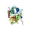
| ||||||||
|---|---|---|---|---|---|---|---|---|---|
| 1 |
| ||||||||
| Unit cell |
|
-Components
| #1: Protein | Mass: 26134.189 Da / Num. of mol.: 1 / Mutation: S195A Source method: isolated from a genetically manipulated source Source: (gene. exp.) Homo sapiens (human) / Tissue: bone marrow / Plasmid: pET24c+ / Production host:  References: UniProt: P49863, Hydrolases; Acting on peptide bonds (peptidases); Serine endopeptidases |
|---|---|
| #2: Water | ChemComp-HOH /  Mass: 18.015 Da / Num. of mol.: 30 / Source method: isolated from a natural source / Formula: H2O Mass: 18.015 Da / Num. of mol.: 30 / Source method: isolated from a natural source / Formula: H2O |
| Has protein modification | Y |
-Experimental details
-Experiment
| Experiment | Method: X-RAY DIFFRACTION / Number of used crystals: 1 |
|---|
- Sample preparation
| Crystal | Density Matthews: 2.04 Å3/Da / Density % sol: 39.73 % | ||||||||||||||||||
|---|---|---|---|---|---|---|---|---|---|---|---|---|---|---|---|---|---|---|---|
| Crystal grow | Temperature: 291 K / Method: vapor diffusion, sitting drop / pH: 8.4 Details: 1.6M sodium formate, 2.5mM 2-[N-morpholino]ethane-sulfonic acid, 50mM sodium chloride, pH 8.4, VAPOR DIFFUSION, SITTING DROP, temperature 291K | ||||||||||||||||||
| Crystal grow | *PLUS | ||||||||||||||||||
| Components of the solutions | *PLUS
|
-Data collection
| Diffraction | Mean temperature: 291 K |
|---|---|
| Diffraction source | Source: ROTATING ANODE / Type: RIGAKU / Wavelength: 1.5418 Å |
| Detector | Type: MARRESEARCH / Detector: IMAGE PLATE / Date: Nov 26, 2000 |
| Radiation | Monochromator: CuK alpha / Protocol: SINGLE WAVELENGTH / Monochromatic (M) / Laue (L): M / Scattering type: x-ray |
| Radiation wavelength | Wavelength: 1.5418 Å / Relative weight: 1 |
| Reflection | Resolution: 2.9→50 Å / Num. all: 39313 / Num. obs: 10537 / % possible obs: 88.8 % / Observed criterion σ(F): 0 / Observed criterion σ(I): 0 / Rmerge(I) obs: 0.147 |
| Reflection shell | Resolution: 2.9→3 Å / Num. unique all: 477 / % possible all: 92.5 |
| Reflection | *PLUS Highest resolution: 2.9 Å / Lowest resolution: 50 Å / % possible obs: 94.5 % / Num. measured all: 39313 |
| Reflection shell | *PLUS % possible obs: 92.5 % |
- Processing
| Software |
| |||||||||||||||||||||||||
|---|---|---|---|---|---|---|---|---|---|---|---|---|---|---|---|---|---|---|---|---|---|---|---|---|---|---|
| Refinement | Method to determine structure: FOURIER SYNTHESIS Starting model: granzyme B Resolution: 2.9→22 Å / Isotropic thermal model: anisotropic / Cross valid method: THROUGHOUT / σ(F): 0 / σ(I): 0 / Stereochemistry target values: Engh & Huber
| |||||||||||||||||||||||||
| Displacement parameters | Biso mean: 26.8 Å2 | |||||||||||||||||||||||||
| Refinement step | Cycle: LAST / Resolution: 2.9→22 Å
| |||||||||||||||||||||||||
| Refine LS restraints |
| |||||||||||||||||||||||||
| Refinement | *PLUS Highest resolution: 2.9 Å / Lowest resolution: 22 Å / Num. reflection obs: 9771 / Num. reflection Rfree: 500 / Rfactor Rfree: 0.3167 / Rfactor Rwork: 0.2236 | |||||||||||||||||||||||||
| Solvent computation | *PLUS | |||||||||||||||||||||||||
| Displacement parameters | *PLUS | |||||||||||||||||||||||||
| Refine LS restraints | *PLUS
|
