 Movie
Movie Controller

[English] 日本語
 Yorodumi
- PDB-1mww: THE STRUCTURE OF THE HYPOTHETICAL PROTEIN HI1388.1 FROM HAEMOPHIL... -
+ Open data

- Basic information
| Entry | Database: PDB / ID: 1mww | ||||||
|---|---|---|---|---|---|---|---|
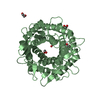
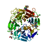
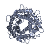
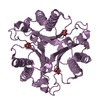
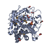
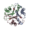
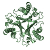
| Title | THE STRUCTURE OF THE HYPOTHETICAL PROTEIN HI1388.1 FROM HAEMOPHILUS INFLUENZAE REVEALS A TAUTOMERASE/MIF FOLD | ||||||
 Components Components | HYPOTHETICAL PROTEIN HI1388.1 | ||||||
 Keywords Keywords | STRUCTURAL GENOMICS / UNKNOWN FUNCTION / I1388.1 / HYPOTHETICAL PROTEIN / Structure 2 Function Project / S2F | ||||||
| Function / homology | Tautomerase, MSAD family / Tautomerase enzyme / Macrophage Migration Inhibitory Factor / Macrophage Migration Inhibitory Factor / Tautomerase/MIF superfamily / 2-Layer Sandwich / Alpha Beta / GLUTAMIC ACID / Uncharacterized protein HI_1388.1 Function and homology information Function and homology information | ||||||
| Biological species |  Haemophilus influenzae (bacteria) Haemophilus influenzae (bacteria) | ||||||
| Method |  X-RAY DIFFRACTION / SYNCHROTRON / MOLECULAR REPLACEMENT / Resolution: 2.08 Å X-RAY DIFFRACTION / SYNCHROTRON / MOLECULAR REPLACEMENT / Resolution: 2.08 Å | ||||||
 Authors Authors | Lehmann, C. / Pullalarevu, S. / Krajewski, W. / Galkin, A. / Howard, A. / Herzberg, O. / Structure 2 Function Project (S2F) | ||||||
 Citation Citation | Journal: To be Published Title: Structure of the Hypothetical Protein HI1388.1 from Haemophilus influenzae Authors: Lehmann, C. / Pullalarevu, S. / Krajewski, W. / Galkin, A. / Howard, A. / Herzberg, O. | ||||||
| History |
|
- Structure visualization
| Structure viewer | Molecule: MolmilJmol/JSmol |
|---|
- Downloads & links
-Download
| PDBx/mmCIF format | 1mww.cif.gz | 91.8 KB | Display | PDBx/mmCIF format |
|---|---|---|---|---|
| PDB format | pdb1mww.ent.gz | 71.4 KB | Display | PDB format |
| PDBx/mmJSON format | 1mww.json.gz | Tree view | PDBx/mmJSON format | |
| Others |  Other downloads Other downloads |
-Validation report
| Arichive directory | https://data.pdbj.org/pub/pdb/validation_reports/mw/1mwwftp://data.pdbj.org/pub/pdb/validation_reports/mw/1mww | HTTPS FTP |
|---|
-Related structure data
| Related structure data | 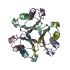 1otfS S: Starting model for refinement |
|---|---|
| Similar structure data | |
| Other databases |


-Links
 PDBj
PDBj

- Assembly
| Deposited unit | 
| ||||||||||||
|---|---|---|---|---|---|---|---|---|---|---|---|---|---|
| 1 |
| ||||||||||||
| Unit cell |
| ||||||||||||
| Components on special symmetry positions |
|
-Components
| #1: Protein | Mass: 14985.425 Da / Num. of mol.: 3 Source method: isolated from a genetically manipulated source Source: (gene. exp.) Haemophilus influenzae (bacteria) / Gene: HI1388.1 / Plasmid: PET100 / Species (production host): Escherichia coli / Production host: #2: Chemical | ChemComp-CL / |   Mass: 35.453 Da / Num. of mol.: 1 / Source method: obtained synthetically / Formula: Cl Mass: 35.453 Da / Num. of mol.: 1 / Source method: obtained synthetically / Formula: Cl#3: Chemical | ChemComp-GLU / |   Type: L-peptide linking / Mass: 147.129 Da / Num. of mol.: 1 / Source method: obtained synthetically / Formula: C5H9NO4 Type: L-peptide linking / Mass: 147.129 Da / Num. of mol.: 1 / Source method: obtained synthetically / Formula: C5H9NO4#4: Water | ChemComp-HOH / |  Mass: 18.015 Da / Num. of mol.: 356 / Source method: isolated from a natural source / Formula: H2O Mass: 18.015 Da / Num. of mol.: 356 / Source method: isolated from a natural source / Formula: H2O |
|---|
-Experimental details
-Experiment
| Experiment | Method: X-RAY DIFFRACTION / Number of used crystals: 1 |
|---|
- Sample preparation
| Crystal | Density Matthews: 2.14 Å3/Da / Density % sol: 46.36 % |
|---|---|
| Crystal grow | Temperature: 294 K / Method: vapor diffusion, sitting drop / pH: 5.5 Details: 50% MPD, 0.1M NACACODYLATE, 0.2M NAGLUTAMATE, pH 5.5, VAPOR DIFFUSION, SITTING DROP, temperature 294K |
-Data collection
| Diffraction | Mean temperature: 100 K |
|---|---|
| Diffraction source | Source: SYNCHROTRON / Site: APS  / Beamline: 17-ID / Wavelength: 0.9724 / Wavelength: 0.9724 Å / Beamline: 17-ID / Wavelength: 0.9724 / Wavelength: 0.9724 Å |
| Detector | Type: ADSC QUANTUM 210 / Detector: CCD / Date: Jul 7, 2002 / Details: MIRROR |
| Radiation | Monochromator: CRYOGENICALLY-COOLED SI(111) DOUBLE-CRYSTAL SYSTEM Protocol: SINGLE WAVELENGTH / Monochromatic (M) / Laue (L): M / Scattering type: x-ray |
| Radiation wavelength | Wavelength: 0.9724 Å / Relative weight: 1 |
| Reflection | Resolution: 2.08→19.83 Å / Num. obs: 24983 / % possible obs: 98.3 % / Observed criterion σ(F): 0 / Observed criterion σ(I): 0 / Redundancy: 14.9 % / Biso Wilson estimate: 29.8 Å2 / Rmerge(I) obs: 0.048 / Net I/σ(I): 15.5 |
| Reflection shell | Resolution: 2.08→2.15 Å / Rmerge(I) obs: 0.305 / Mean I/σ(I) obs: 8.96 / Num. unique all: 24986 / % possible all: 89.6 |
- Processing
| Software |
| ||||||||||||||||||||
|---|---|---|---|---|---|---|---|---|---|---|---|---|---|---|---|---|---|---|---|---|---|
| Refinement | Method to determine structure: MOLECULAR REPLACEMENT Starting model: 1OTF Resolution: 2.08→19.83 Å / Cross valid method: THROUGHOUT / σ(F): 0 / Stereochemistry target values: Engh & Huber
| ||||||||||||||||||||
| Solvent computation | Solvent model: MOEWS & KRETSINGER, J.MOL.BIOL.91(1973)201-228 | ||||||||||||||||||||
| Displacement parameters | Biso mean: 32.6 Å2 | ||||||||||||||||||||
| Refinement step | Cycle: LAST / Resolution: 2.08→19.83 Å
| ||||||||||||||||||||
| Refine LS restraints |
| ||||||||||||||||||||
| LS refinement shell | Resolution: 2.08→2.17 Å
|
