 Movie
Movie Controller

[English] 日本語
 Yorodumi
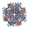
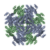
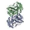
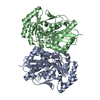
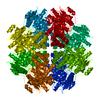
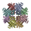
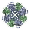
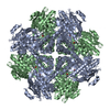
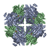
- PDB-1muc: STRUCTURE OF MUCONATE LACTONIZING ENZYME AT 1.85 ANGSTROMS RESOLUTION -
+ Open data

- Basic information
| Entry | Database: PDB / ID: 1muc | |||||||||
|---|---|---|---|---|---|---|---|---|---|---|
| Title | STRUCTURE OF MUCONATE LACTONIZING ENZYME AT 1.85 ANGSTROMS RESOLUTION | |||||||||
 Components Components | MUCONATE LACTONIZING ENZYME | |||||||||
 Keywords Keywords | ISOMERASE / MUCONATE LACTONIZING ENZYME | |||||||||
| Function / homology |  Function and homology information Function and homology informationmuconate cycloisomerase / chloromuconate cycloisomerase activity / muconate cycloisomerase activity / racemase and epimerase activity / beta-ketoadipate pathway / amino acid catabolic process / peptide metabolic process / manganese ion binding Similarity search - Function | |||||||||
| Biological species |  Pseudomonas putida (bacteria) Pseudomonas putida (bacteria) | |||||||||
| Method |  X-RAY DIFFRACTION / Resolution: 1.85 Å X-RAY DIFFRACTION / Resolution: 1.85 Å | |||||||||
 Authors Authors | Helin, S. / Kahn, P.C. / Guha, B.H.L. / Mallows, D.J. / Goldman, A. | |||||||||
 Citation Citation | Journal: J.Mol.Biol. / Year: 1995 Title: The refined X-ray structure of muconate lactonizing enzyme from Pseudomonas putida PRS2000 at 1.85 A resolution. Authors: Helin, S. / Kahn, P.C. / Guha, B.L. / Mallows, D.G. / Goldman, A. #1: Journal: J.Mol.Biol. / Year: 1987Title: Crystal Structure of Muconate Lactonizing Enzyme at 3 A Resolution Authors: Goldman, A. / Ollis, D.L. / Steitz, T.A. #2: Journal: J.Mol.Biol. / Year: 1985Title: Crystal Structure of Muconate Lactonizing Enzyme at 6.5 A Resolution Authors: Goldman, A. / Ollis, D. / Ngai, K.L. / Steitz, T.A. | |||||||||
| History |
|
- Structure visualization
| Structure viewer | Molecule: MolmilJmol/JSmol |
|---|
- Downloads & links
-Download
| PDBx/mmCIF format | 1muc.cif.gz | 151 KB | Display | PDBx/mmCIF format |
|---|---|---|---|---|
| PDB format | pdb1muc.ent.gz | 118.6 KB | Display | PDB format |
| PDBx/mmJSON format | 1muc.json.gz | Tree view | PDBx/mmJSON format | |
| Others |  Other downloads Other downloads |
-Validation report
| Arichive directory | https://data.pdbj.org/pub/pdb/validation_reports/mu/1mucftp://data.pdbj.org/pub/pdb/validation_reports/mu/1muc | HTTPS FTP |
|---|
-Related structure data
| Similar structure data |
|---|

-Links
 PDBj
PDBj
- Assembly
| Deposited unit | 
| ||||||||
|---|---|---|---|---|---|---|---|---|---|
| 1 | 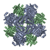
| ||||||||
| Unit cell |
| ||||||||
| Noncrystallographic symmetry (NCS) | NCS oper: (Code: given Matrix: (0.7537, 0.6572, 0.0035), Vector: |
-Components
| #1: Protein | Mass: 40355.172 Da / Num. of mol.: 2 / Source method: isolated from a natural source / Source: (natural) Pseudomonas putida (bacteria) / Strain: PRS2000 / References: UniProt: P08310, muconate cycloisomerase#2: Chemical |   Mass: 54.938 Da / Num. of mol.: 2 / Source method: obtained synthetically / Formula: Mn Mass: 54.938 Da / Num. of mol.: 2 / Source method: obtained synthetically / Formula: Mn#3: Water | ChemComp-HOH / |  Mass: 18.015 Da / Num. of mol.: 307 / Source method: isolated from a natural source / Formula: H2O Mass: 18.015 Da / Num. of mol.: 307 / Source method: isolated from a natural source / Formula: H2O |
|---|
-Experimental details
-Experiment
| Experiment | Method: X-RAY DIFFRACTION / Number of used crystals: 4 |
|---|
- Sample preparation
| Crystal | Density Matthews: 2.53 Å3/Da / Density % sol: 52 % |
|---|---|
| Crystal | *PLUS |
| Crystal grow | *PLUS Method: unknown |
-Data collection
| Diffraction source | Wavelength: 1.5418 |
|---|---|
| Detector | Type: XUONG-HAMLIN MULTIWIRE / Detector: AREA DETECTOR / Date: Jan 1, 1985 |
| Radiation | Monochromatic (M) / Laue (L): M / Scattering type: x-ray |
| Radiation wavelength | Wavelength: 1.5418 Å / Relative weight: 1 |
| Reflection | Num. obs: 66609 / % possible obs: 87.2 % / Redundancy: 3.2 % / Rmerge(I) obs: 0.093 |
| Reflection | *PLUS Highest resolution: 1.85 Å / Lowest resolution: 8 Å / Num. obs: 59010 / Num. measured all: 191642 / Rmerge(I) obs: 0.193 |
- Processing
| Software |
| ||||||||||||||||||||||||||||||||||||||||||||||||||||||||||||
|---|---|---|---|---|---|---|---|---|---|---|---|---|---|---|---|---|---|---|---|---|---|---|---|---|---|---|---|---|---|---|---|---|---|---|---|---|---|---|---|---|---|---|---|---|---|---|---|---|---|---|---|---|---|---|---|---|---|---|---|---|---|
| Refinement | Resolution: 1.85→8 Å / σ(F): 1 Details: FOR RESIDUE ARG A 20 ELECTRON DENSITY WAS VISIBLE ONLY FOR BACKBONE ATOMS.
| ||||||||||||||||||||||||||||||||||||||||||||||||||||||||||||
| Displacement parameters | Biso mean: 21 Å2 | ||||||||||||||||||||||||||||||||||||||||||||||||||||||||||||
| Refine analyze | Luzzati coordinate error obs: 0.2 Å | ||||||||||||||||||||||||||||||||||||||||||||||||||||||||||||
| Refinement step | Cycle: LAST / Resolution: 1.85→8 Å
| ||||||||||||||||||||||||||||||||||||||||||||||||||||||||||||
| Refine LS restraints |
| ||||||||||||||||||||||||||||||||||||||||||||||||||||||||||||
| Software | *PLUS Name: X-PLOR / Version: 3.1 / Classification: refinement | ||||||||||||||||||||||||||||||||||||||||||||||||||||||||||||
| Refine LS restraints | *PLUS
|
