 Movie
Movie Controller


+ Open data

- Basic information
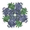
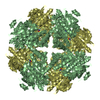
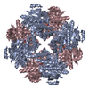
| Entry | Database: PDB / ID: 1f9c | ||||||
|---|---|---|---|---|---|---|---|
| Title | CRYSTAL STRUCTURE OF MLE D178N VARIANT | ||||||
 Components Components | PROTEIN (MUCONATE CYCLOISOMERASE I) | ||||||
 Keywords Keywords | ISOMERASE / THERMOSTABLE MUTANT | ||||||
| Function / homology |  Function and homology information Function and homology informationchloromuconate cycloisomerase activity / muconate cycloisomerase activity / racemase and epimerase activity / amino acid catabolic process / peptide metabolic process / manganese ion binding Similarity search - Function | ||||||
| Biological species |  Pseudomonas putida (bacteria) Pseudomonas putida (bacteria) | ||||||
| Method |  X-RAY DIFFRACTION / SYNCHROTRON / Resolution: 2.5 Å X-RAY DIFFRACTION / SYNCHROTRON / Resolution: 2.5 Å | ||||||
 Authors Authors | Kajander, T. / Lehtio, L. / Kahn, P.C. / Goldman, A. | ||||||
 Citation Citation | Journal: Structure Fold.Des. / Year: 2000 Title: Buried charged surface in proteins. Authors: Kajander, T. / Kahn, P.C. / Passila, S.H. / Cohen, D.C. / Lehtio, L. / Adolfsen, W. / Warwicker, J. / Schell, U. / Goldman, A. #1: Journal: J.Mol.Biol. / Year: 1995Title: The Refined Crystal Structure of Muconate Lactonising Enzyme from Pseudomonas putida PRS2000 at 1.85 Resolution Authors: Helin, S. / Kahn, P.C. / Guha, B.L. / Mallows, D.G. / Goldman, A. | ||||||
| History |
|
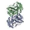
- Structure visualization
| Structure viewer | Molecule: MolmilJmol/JSmol |
|---|
- Downloads & links
-Download
| PDBx/mmCIF format | 1f9c.cif.gz | 137.5 KB | Display | PDBx/mmCIF format |
|---|---|---|---|---|
| PDB format | pdb1f9c.ent.gz | 109.4 KB | Display | PDB format |
| PDBx/mmJSON format | 1f9c.json.gz | Tree view | PDBx/mmJSON format | |
| Others |  Other downloads Other downloads |
-Validation report
| Arichive directory | https://data.pdbj.org/pub/pdb/validation_reports/f9/1f9cftp://data.pdbj.org/pub/pdb/validation_reports/f9/1f9c | HTTPS FTP |
|---|
-Related structure data
| Related structure data | |
|---|---|
| Similar structure data |


-Links
 PDBj
PDBj
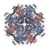
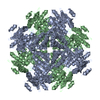
- Assembly
| Deposited unit | 
| ||||||||
|---|---|---|---|---|---|---|---|---|---|
| 1 | 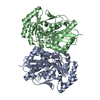
| ||||||||
| Unit cell |
|
-Components
| #1: Protein | Mass: 40107.832 Da / Num. of mol.: 2 / Mutation: YES Source method: isolated from a genetically manipulated source Source: (gene. exp.) Pseudomonas putida (bacteria) / Strain: PRS2000 / Plasmid: PET11A / Production host: #2: Chemical |   Mass: 54.938 Da / Num. of mol.: 2 / Source method: obtained synthetically / Formula: Mn Mass: 54.938 Da / Num. of mol.: 2 / Source method: obtained synthetically / Formula: Mn#3: Water | ChemComp-HOH / |  Mass: 18.015 Da / Num. of mol.: 195 / Source method: isolated from a natural source / Formula: H2O Mass: 18.015 Da / Num. of mol.: 195 / Source method: isolated from a natural source / Formula: H2O |
|---|
-Experimental details
-Experiment
| Experiment | Method: X-RAY DIFFRACTION / Number of used crystals: 1 |
|---|
- Sample preparation
| Crystal | Density Matthews: 2.42 Å3/Da / Density % sol: 49.28 % |
|---|---|
| Crystal grow | Temperature: 277 K / Method: microdialysis / pH: 6.5 Details: MES, NaCl, MnCl2, beta-mercaptoethanol, NaN3, pH 6.5, MICRODIALYSIS, temperature 277K |
-Data collection
| Diffraction | Mean temperature: 103 K |
|---|---|
| Diffraction source | Source: SYNCHROTRON / Site: EMBL/DESY, HAMBURG  / Beamline: X31 / Wavelength: 0.92 / Beamline: X31 / Wavelength: 0.92 |
| Detector | Type: MARRESEARCH / Detector: IMAGE PLATE / Date: Sep 1, 1998 |
| Radiation | Protocol: SINGLE WAVELENGTH / Monochromatic (M) / Laue (L): M / Scattering type: x-ray |
| Radiation wavelength | Wavelength: 0.92 Å / Relative weight: 1 |
| Reflection | Resolution: 2.5→20 Å / Num. obs: 22444 / % possible obs: 87 % / Rmerge(I) obs: 0.046 |
| Reflection | *PLUS Highest resolution: 2.5 Å / Lowest resolution: 20 Å |
| Reflection shell | *PLUS Highest resolution: 2.5 Å / Lowest resolution: 2.6 Å / % possible obs: 59.3 % |
- Processing
| Software |
| ||||||||||||||||||||||||||||||||||||||||||||||||||||||||||||
|---|---|---|---|---|---|---|---|---|---|---|---|---|---|---|---|---|---|---|---|---|---|---|---|---|---|---|---|---|---|---|---|---|---|---|---|---|---|---|---|---|---|---|---|---|---|---|---|---|---|---|---|---|---|---|---|---|---|---|---|---|---|
| Refinement | Resolution: 2.5→8 Å / σ(F): 2
| ||||||||||||||||||||||||||||||||||||||||||||||||||||||||||||
| Refinement step | Cycle: LAST / Resolution: 2.5→8 Å
| ||||||||||||||||||||||||||||||||||||||||||||||||||||||||||||
| Refine LS restraints |
| ||||||||||||||||||||||||||||||||||||||||||||||||||||||||||||
| Software | *PLUS Name: X-PLOR / Version: 3.851 / Classification: refinement | ||||||||||||||||||||||||||||||||||||||||||||||||||||||||||||
| Refinement | *PLUS % reflection Rfree: 5 % / Rfactor all: 0.187 / Rfactor obs: 0.18 | ||||||||||||||||||||||||||||||||||||||||||||||||||||||||||||
| Solvent computation | *PLUS | ||||||||||||||||||||||||||||||||||||||||||||||||||||||||||||
| Displacement parameters | *PLUS | ||||||||||||||||||||||||||||||||||||||||||||||||||||||||||||
| Refine LS restraints | *PLUS
|
