 Movie
Movie Controller


+ Open data

- Basic information
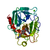
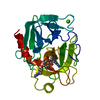
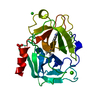
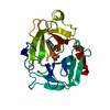
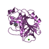
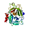
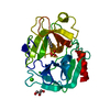
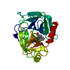
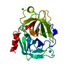
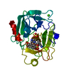
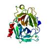
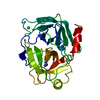
| Entry | Database: PDB / ID: 1mtw | ||||||
|---|---|---|---|---|---|---|---|
| Title | FACTOR XA SPECIFIC INHIBITOR IN COMPLEX WITH BOVINE TRYPSIN | ||||||
 Components Components | TRYPSIN | ||||||
 Keywords Keywords | SERINE PROTEASE / HYDROLASE | ||||||
| Function / homology |  Function and homology information Function and homology informationtrypsin / serpin family protein binding / serine protease inhibitor complex / digestion / endopeptidase activity / serine-type endopeptidase activity / proteolysis / extracellular space / metal ion binding Similarity search - Function | ||||||
| Biological species |  | ||||||
| Method |  X-RAY DIFFRACTION / DIFFERENCE FOURIER / Resolution: 1.9 Å X-RAY DIFFRACTION / DIFFERENCE FOURIER / Resolution: 1.9 Å | ||||||
 Authors Authors | Stubbs, M.T. | ||||||
 Citation Citation | Journal: FEBS Lett. / Year: 1995 Title: Crystal structures of factor Xa specific inhibitors in complex with trypsin: structural grounds for inhibition of factor Xa and selectivity against thrombin. Authors: Stubbs, M.T. / Huber, R. / Bode, W. #1: Journal: J.Mol.Biol. / Year: 1997Title: The Second Kunitz Domain of Human Tissue Factor Pathway Inhibitor. Cloning, Structure Determination and Interaction with Factor Xa Authors: Burgering, M.J. / Orbons, L.P. / Van Der Doelen, A. / Mulders, J. / Theunissen, H.J. / Grootenhuis, P.D. / Bode, W. / Huber, R. / Stubbs, M.T. #2: Journal: Embo J. / Year: 1996Title: The Ornithodorin-Thrombin Crystal Structure, a Key to the Tap Enigma? Authors: Van De Locht, A. / Stubbs, M.T. / Bode, W. / Friedrich, T. / Bollschweiler, C. / Hoffken, W. / Huber, R. #3: Journal: Curr.Pharm.Des. / Year: 1996Title: Structural Aspects of Factor Xa Inhibition Authors: Stubbs, M.T. #4: Journal: J.Biol.Chem. / Year: 1996Title: X-Ray Structure of Active Site-Inhibited Clotting Factor Xa. Implications for Drug Design and Substrate Recognition Authors: Brandstetter, H. / Kuhne, A. / Bode, W. / Huber, R. / Von Der Saal, W. / Wirthensohn, K. / Engh, R.A. #5: Journal: J.Mol.Biol. / Year: 1993Title: Structure of Human Des(1-45) Factor Xa at 2.2 A Resolution Authors: Padmanabhan, K. / Padmanabhan, K.P. / Tulinsky, A. / Park, C.H. / Bode, W. / Huber, R. / Blankenship, D.T. / Cardin, A.D. / Kisiel, W. #6: Journal: FEBS Lett. / Year: 1991Title: Geometry of Binding of the N Alpha-Tosylated Piperidides of M-Amidino-, P-Amidino-and P-Guanidino Phenylalanine to Thrombin and Trypsin. X-Ray Crystal Structures of Their Trypsin Complexes and ...Title: Geometry of Binding of the N Alpha-Tosylated Piperidides of M-Amidino-, P-Amidino-and P-Guanidino Phenylalanine to Thrombin and Trypsin. X-Ray Crystal Structures of Their Trypsin Complexes and Modeling of Their Thrombin Complexes Authors: Turk, D. / Sturzebecher, J. / Bode, W. #7: Journal: Eur.J.Biochem. / Year: 1990Title: Geometry of Binding of the Benzamidine-and Arginine-Based Inhibitors N Alpha-(2-Naphthyl-Sulphonyl-Glycyl)-Dl-P-Amidinophenylalanyl-Piperidine (Napap) and (2R,4R)-4-Methyl-1-[N Alpha-(3-Methyl- ...Title: Geometry of Binding of the Benzamidine-and Arginine-Based Inhibitors N Alpha-(2-Naphthyl-Sulphonyl-Glycyl)-Dl-P-Amidinophenylalanyl-Piperidine (Napap) and (2R,4R)-4-Methyl-1-[N Alpha-(3-Methyl-1,2,3,4-Tetrahydro-8-Quinolinesulphonyl)-L-Arginyl]-2-Piperidine to Carboxylic Acid (Mqpa) Human Alpha-Thrombin. X-Ray Crystallographic Determination of the Napap-Trypsin Complex and Modeling of Napap-Thrombin and Mqpa-Thrombin Authors: Bode, W. / Turk, D. / Sturzebecher, J. #8: Journal: J.Mol.Biol. / Year: 1989Title: Crystal Structure of Bovine Beta-Trypsin at 1.5 A Resolution in a Crystal Form with Low Molecular Packing Density. Active Site Geometry, Ion Pairs and Solvent Structure Authors: Bartunik, H.D. / Summers, L.J. / Bartsch, H.H. #9: Journal: J.Mol.Biol. / Year: 1975Title: The Refined Crystal Structure of Bovine Beta-Trypsin at 1.8 A Resolution. II. Crystallographic Refinement, Calcium Binding Site, Benzamidine Binding Site and Active Site at Ph 7.0 Authors: Bode, W. / Schwager, P. | ||||||
| History |
|
- Structure visualization
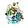
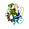
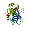
| Structure viewer | Molecule: MolmilJmol/JSmol |
|---|
- Downloads & links
-Download
| PDBx/mmCIF format | 1mtw.cif.gz | 63.1 KB | Display | PDBx/mmCIF format |
|---|---|---|---|---|
| PDB format | pdb1mtw.ent.gz | 44.6 KB | Display | PDB format |
| PDBx/mmJSON format | 1mtw.json.gz | Tree view | PDBx/mmJSON format | |
| Others |  Other downloads Other downloads |
-Validation report
| Summary document | 1mtw_validation.pdf.gz | 734.1 KB | Display | wwPDB validaton report |
|---|---|---|---|---|
| Full document | 1mtw_full_validation.pdf.gz | 736.3 KB | Display | |
| Data in XML | 1mtw_validation.xml.gz | 15.5 KB | Display | |
| Data in CIF | 1mtw_validation.cif.gz | 22.4 KB | Display | |
| Arichive directory | https://data.pdbj.org/pub/pdb/validation_reports/mt/1mtwftp://data.pdbj.org/pub/pdb/validation_reports/mt/1mtw | HTTPS FTP |
-Related structure data


-Links
 PDBj
PDBj

- Assembly
| Deposited unit | 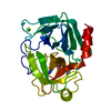
| ||||||||
|---|---|---|---|---|---|---|---|---|---|
| 1 |
| ||||||||
| Unit cell |
|
-Components
| #1: Protein | Mass: 23324.287 Da / Num. of mol.: 1 / Source method: isolated from a natural source / Source: (natural) |
|---|---|
| #2: Chemical | ChemComp-CA /   Mass: 40.078 Da / Num. of mol.: 1 / Source method: obtained synthetically / Formula: Ca Mass: 40.078 Da / Num. of mol.: 1 / Source method: obtained synthetically / Formula: Ca |
| #3: Chemical | ChemComp-DX9 / (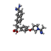  Mass: 444.526 Da / Num. of mol.: 1 / Source method: obtained synthetically / Formula: C26H28N4O3 Mass: 444.526 Da / Num. of mol.: 1 / Source method: obtained synthetically / Formula: C26H28N4O3 |
| #4: Water | ChemComp-HOH /  Mass: 18.015 Da / Num. of mol.: 289 / Source method: isolated from a natural source / Formula: H2O Mass: 18.015 Da / Num. of mol.: 289 / Source method: isolated from a natural source / Formula: H2O |
| Has protein modification | Y |
-Experimental details
-Experiment
| Experiment | Method: X-RAY DIFFRACTION / Number of used crystals: 1 |
|---|
- Sample preparation
| Crystal | Density Matthews: 2.31 Å3/Da / Density % sol: 46.69 % | |||||||||||||||
|---|---|---|---|---|---|---|---|---|---|---|---|---|---|---|---|---|
| Crystal grow | pH: 8 / Details: 0.3M AMMONIUM SULFATE, PH 8.0, 30% PEG 8000 | |||||||||||||||
| Crystal grow | *PLUS Method: vapor diffusion | |||||||||||||||
| Components of the solutions | *PLUS
|
-Data collection
| Diffraction | Mean temperature: 287 K |
|---|---|
| Diffraction source | Source: ROTATING ANODE / Type: RIGAKU / Wavelength: 1.5418 |
| Detector | Type: SIEMENS / Detector: AREA DETECTOR / Date: Nov 1, 1994 / Details: MIRRORS |
| Radiation | Monochromator: NI FILTER / Monochromatic (M) / Laue (L): M / Scattering type: x-ray |
| Radiation wavelength | Wavelength: 1.5418 Å / Relative weight: 1 |
| Reflection | Resolution: 1.9→20 Å / Num. obs: 16195 / % possible obs: 91.7 % / Observed criterion σ(I): 2 / Redundancy: 1.6 % / Rmerge(I) obs: 0.032 / Rsym value: 0.032 / Net I/σ(I): 12 |
| Reflection shell | Resolution: 1.9→2 Å / Redundancy: 1.3 % / Rmerge(I) obs: 0.119 / Mean I/σ(I) obs: 3 / Rsym value: 0.119 / % possible all: 80.3 |
| Reflection | *PLUS Num. measured all: 25539 |
- Processing
| Software |
| ||||||||||||||||||||||||||||||||||||||||||||||||||||||||||||
|---|---|---|---|---|---|---|---|---|---|---|---|---|---|---|---|---|---|---|---|---|---|---|---|---|---|---|---|---|---|---|---|---|---|---|---|---|---|---|---|---|---|---|---|---|---|---|---|---|---|---|---|---|---|---|---|---|---|---|---|---|---|
| Refinement | Method to determine structure: DIFFERENCE FOURIER / Resolution: 1.9→8 Å / Data cutoff high absF: 10000000 / Data cutoff low absF: 0.001 / σ(F): 3
| ||||||||||||||||||||||||||||||||||||||||||||||||||||||||||||
| Refine analyze | Luzzati d res low obs: 8 Å | ||||||||||||||||||||||||||||||||||||||||||||||||||||||||||||
| Refinement step | Cycle: LAST / Resolution: 1.9→8 Å
| ||||||||||||||||||||||||||||||||||||||||||||||||||||||||||||
| Refine LS restraints |
| ||||||||||||||||||||||||||||||||||||||||||||||||||||||||||||
| LS refinement shell | Resolution: 1.9→1.93 Å / Total num. of bins used: 20
| ||||||||||||||||||||||||||||||||||||||||||||||||||||||||||||
| Xplor file |
| ||||||||||||||||||||||||||||||||||||||||||||||||||||||||||||
| Software | *PLUS Name: X-PLOR / Version: 3.1 / Classification: refinement | ||||||||||||||||||||||||||||||||||||||||||||||||||||||||||||
| Refine LS restraints | *PLUS
| ||||||||||||||||||||||||||||||||||||||||||||||||||||||||||||
| LS refinement shell | *PLUS Rfactor Rwork: 0.31 |
